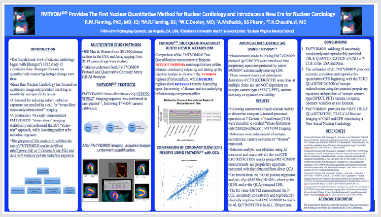
FMTVDM©℗ Provides the First Patented Quantification of Myocardial Perfusion Imaging (MPI) Yielding Theranostic Benefit for Individuals with Suspected Coronary Artery Disease (CAD)
Introduction
Myocardial perfusion imaging (MPI) using either single photon emission computed tomography (SPECT)/Planar or positron emission tomography (PET) has been limited by the utilization of qualitative image interpretation. This qualitative approach has restricted the use of SPECT/Planar and PET to assessment of coronary artery disease (CAD) as either present (sensitivity) or absent (specificity). Consequently, determination of treatment success has been constrained. The introduction of the first truly quantitative method [1-8] for measuring CAD and regional blood flow differences (RBFDs), providing artificial intelligence (AI)/machine learning (ML) application has been compared with conventional qualitative imaging [9-10] and was recently demonstrated at the 2018 American Society of Nuclear Cardiology [11] Conference. The clinical significance and application of this method for BOTH diagnostic assessment and treatment monitoring and adjustment (viz. theranostics) is shown here in two cases. The first where conventional MPI missed critical CAD (lack of sensitivity), which would have cost the veteran his life had the critical disease not been caught by FMTVDM and the second where conventional MPI resulted in invasive coronary angiography, which produced adverse vascular consequences resulting in the patient being placed on a balloon pump (lack of specificity).
Case One
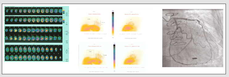
FMTVDM finds critical CAD missed using qualitative MPI. A late middle-aged Caucasian male presented to his community hospital for evaluation of intermittent retrosternal chest discomfort. He was scheduled for diagnostic evaluation comparing [9-12] qualitative MPI imaging with quantitative FMTVDM (Figure 1). Figure 2a shows conventional MPI qualitative image display with and without attenuation correction. The clinician/physician interpretation of the qualitative study was “no evidence of ischemic CAD”. Figure 2b shows the results displayed quantitatively, in vertical and longaxis views. FMTVDM quantification showed wash-in of the isotope (redistribution) demonstrating critical anterior, anteroseptal and anterolateral CAD. The patient was referred for coronary angiography and PCI (Figure 2c) treatment based upon MPI quantitative analysis.
Note: Sequential FMTVDM quantification of isotope redistribution, measuring metabolic and regional blood flow differences (RBFDs).
Figure 1: True FMTVDM Quantification of Myocardial Perfusion Imaging and RBFDs.

a) Standard display of qualitative imaging including attenuation correction (AC images), reveal no evidence of CAD.
(b) Quantification of the patients MPI demonstrates isotope redistribution, with the left panel demonstrating isotope measurement at 5-minutes post stress and the right panel demonstrating 60-minutes post stress imaging.
(c) The quantified images resulted in percutaneous coronary intervention (PCI) of the left anterior descending (LAD), 1st diagonal and 1st obtuse marginal arteries as demonstrated in the before (left) and after (right) panels.
Figure 2: Critical CAD missed using conventional qualitative MPI.

Case Two
FMTVDM quantification demonstrates patent coronary artery bypass graft (CABG) without RBFDs/CAD. A late middle-aged Caucasian female presented with dyspnea on exertion and a history of CAD previously treated by CABG. Her physician ordered a “stress-rest” qualitative MPI study. She also underwent FMTVDM quantitative imaging [9-12] using the same protocol [1-6, 8-11] employed in case one (Figure 1). Physician interpretation of her qualitative MPI (Figure 3a) reported multivessel CAD, involving the LAD and circumflex marginal system. Quantitative FMTVDM measurement (Figure 3b) revealed no evidence of coronary ischemia, which was confirmed angiographically (Figure 3c) with a patent left internal mammary artery (LIMA) graft to her 1st diagonal (D1), 1st obtuse marginal (OM1) and posterior descending (PDA) arteries. During the course of the patient’s coronary angiogram, vascular complications resulted from the procedure and she ended upon on an intra-aortic balloon pump (IABP) for several days in the coronary care unit. She made a complete recovery without further coronary artery intervention or treatment.
(a) Standard display of qualitative imaging was interpreted as showing multi vessel LAD and circumflex CAD.
(b) Quantification of patients MPI revealed no evidence of CAD with a total 5-minute measurement (upper left) of 280,026 and a 60-minute measurement (lower left) of 236,089, revealing a washout of 5.7%, excluding ischemic CAD.
(c) Coronary angiography reveals no evidence of ischemia, with patent LIMA bypass graft to her 1st diagonal, 1st obtuse marginal and posterior descending arteries.
Figure 3: Qualitative MPI interpreted as demonstrating ischemia, while quantitative measurement revealed no ischemia.

Discussion
Limitations in qualitative assessment of disease [12] including assessment of coronary artery disease from MPI and coronary arteriography [13] has resulted in the call for the development of quantitative methods to accurately measure [1-8] outcomes to provide for better clinical care and management of patients. Documentation of the diagnostic benefit of this FMTVDM has been well established [9-11]. These two case studies demonstrate the needed benefit of using FMTVDM quantitative imaging to better manage patient diagnostics, treatment and clinical outcomes. The first example revealed the importance of using FMTVDM to find CAD in a patient whose critically narrowed coronary arteries were missed using two-injection “stress-rest” qualitative imaging; the wrong imaging sequence promulgated by misrepresentations made by the pharmaceutical company to the FDA regarding Sestamibi redistribution [3,5,6,8]. In the second case a woman who had previously undergone successful left internal mammary artery (LIMA) bypass surgery, demonstrated complete patency and regional blood flow through the LIMA graft to her native coronary arteries, quantitatively measured by FMTVDM imaging, while the conventional “stress-rest” two injection method promulgated by the same pharmaceutical company and the resultant physician qualitative interpretations, indicated a need for further invasive testing and intervention, which in the end was not needed and consequently resulted in the rare but statistically recognized interventional vascular complications.
Conclusion
The introduction of FMTVDM quantitative MPI provides enhanced patient management and treatment superior to qualitative image interpretation of MPI, using the first patented truly AI quantitative measurement of CAD and regional blood flow differences (RBFDs) to initially define the extent of CAD and then to measure the truly quantitative affect of patient-specific, patientoriented treatment; saving time, money and lives. COI: FMTVDM; B.E.S.T. Imaging©℗ patent is issued to the Primary Author.
Identification and Isolation of the Yeasts in Traditional Yogurts Collected From Villages in Gaziantep,Turkey-https://biomedres01.blogspot.com/2021/01/identification-and-isolation-of-yeasts_30.html
More BJSTR Articles : https://biomedres01.blogspot.com

No comments:
Post a Comment
Note: Only a member of this blog may post a comment.