DEXA Prototype Using SrI2:Eu2+ Coupled to Silicon Photomultiplier
Osteoporosis is a bone disease characterized by low Bone Mineral Density (BMD) leading to a reduction in bone quality. Up to 1 in 2 women and 1 in 4 men over the age of 50 will endure osteoporosis related fractures, making the disease a public health threat [1]. Dual-energy X-ray absorptiometry (DEXA) is the gold standard technique to measure BMD and diagnose osteoporosis. DEXA uses a small dose of ionizing radiation to create images of bones and to measure BMD. Although DEXA’s ability to reproduce BMD measurements is commended, there are some limitations to its technology causing its results to be misleading. Faulty interpretation of DEXA results by physicians leads to an over estimation of BMD in people with large bones and an underestimation of BMD in people with smaller bones [2]. Interpretation leading to misdiagnosis leads to increased medical expenses. In 2017, the National Osteoporosis Foundation reported that osteoporosis related fractures costs are responsible for up to $19 billion annually to patents and the healthcare system [1]. By 2025, experts project the cost to increase to $25 billion annually [1]. With an increased awareness of osteoporosis and the inaccurate assessments of BMD by DEXA, we aim to tackle DEXA’s lack of accuracy in determining BMD by improving and refining materials used as a gamma ray detector. We have developed a DEXA prototype that couples a SrI2 :Eu2+ scintillator with a SiPM. SrI2 :Eu2+ scintillator is a novel material developed at Fisk University. The SiPM is a low power, high efficiency photo-sensor that can offer the capabilities to develop imagers with better spatial resolution than Photomultiplier Tube (PMT).
The optimization of the DEXA detector will lower the cost of the scanner and enhance its performance for proper BMD reports. Europium activated strontium iodide was discovered to be a highperformance scintillator at room temperature in 2007. Cherepy et al. [3] reported on its scintillation properties for crystals grown by the Bridgman method showing a light yield of 90,000 photons/ MeV. SrI2 :Eu2+ shows promise for high energy resolution, with a reported FWHM energy resolution at 662 keV of 2.5% close to the 2% of the semiconductor CZT [4,5]. The price for CZT detectors is considerably high due to raw material processing and defect-free growth difficulties. The segregation coefficient of Zn along the growth axis in CZT is large (k = 1.35) leading to the high compositional variation [6]. SrI2 :Eu2+ scintillation emission spectrum presents a sharp peak around 435 nm which is in the maximum photon detection efficiency range of the SiPM produced by SensL that will be used in the experiments [3,7]. The low power consumption and the small size of SiPM creates the premises to reduce the size of the device and make it portable.
Experimental Methods
A 4 x 4 array composed of 3 mm x 3 mm x 8 mm SrI2:Eu2+ crystals (Figure 1a) were encapsulated in an aluminum package hermitized with a quartz window (Figure 1b). Each crystal of the array was surrounded by GORE reflector on all but one side in order to redirect all the scintillation light toward the SiPM (Figure 1c).
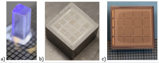
Figure 1:
a. A 3x3x8 mm3 SrI2 :Eu2+ crystal
b. The encapsulated SrI2 :Eu2+ array
c. A SensL 4x4 SiPM array.
The 16 crystals of SrI2 :Eu2+ array are aligned with the 16 pixels (3x3 mm) of SiPM. All the scintillation light produced by the interaction of ionizing radiation with a SrI2 :Eu2+ crystal will be converted by the respective SiPM pixel into an electrical signal. A cylindrical bone phantom (1.5 cm thick) containing CaHPO4 as substituent of bone and resin as soft tissue was prepared. Two other samples composed of aluminum (2.3 mm thick) and copper (0.2 mm thick) were also used into the experiments. The density of aluminum (ρ = 2.7 g/cm3 ) is comparable to that of CaHPO4 (ρ = 2.89 g/cm3 ), while copper is much denser (= 8.96 g/cm3 ) [8]. Table 1 presents the size and the theoretical mass-thickness values of each sample used in the experiments Theoretical mass-thickness, x = ρ t, is calculated using the thickness of each sample and their respective densities. In the case of bone phantom the mass thickness (BMD) was determined taking in consideration the volume fraction (0.1) occupied by the CaHPO4 in the mixture with resin.
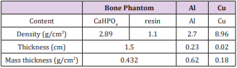
Table 1: Parameters of the samples used in theoretical and experimental determination of mass thickness.
Figure 2 presents the schematic of the setup used in our experiments. The 4 x 4 array of 3 mm x 3 mm x 8 mm SrI2 :Eu2+ crystals encapsulated in an Al package sits on top the SiPM Array SB-4 from SensL. Using the output signal of SiPM processed by the preamplifier and a Pixie 16 Multi-Channel Analyzer (MCA), radiation spectra are acquired for each sample placed on top of the packaged scintillator. The two energies (241Am - 60 keV and 57Co - 122 keV) are attenuated differently by each sample. The spectrum obtained with a sample will provide data about count rate, I, at 60 keV and at 122 keV. A separate spectrum is acquired without any sample covering the detector which will serve as a measure of incoming count rate I0 .The transport of the beams is described by two similar equations: [9]
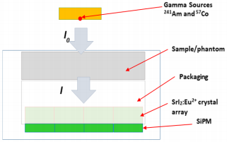
Figure 2: Schematic of an imager using SrI2 :Eu2+ coupled with SiPM.

where I is the count rate measured through the attenuating material, I 0 is the count rate measured without the attenuating material, µ/ρ is the mass attenuation coefficient, x = ρ t is the massthickness or BMD value, ρ is density of attenuating material and t is thickness of attenuating material. In both equation b and s are referring to bone and soft tissue, respectively. The 60 and 122 indexes are referring to the two energies (241Am - 60 keV and 57Co - 122 keV) used in the experiments. Solving the system of equations for xb results in the following:
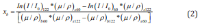
Using Eq 2, experimental value of BMD will be determined. Aluminum and Copper samples served as reference in terms of densities to validate the performance of the system. Since no soft tissue substituent is present Eq 1 and Eq 2 will be simplified as following:
Results
The experimental value of attenuation I/I0 caused by each material is compared with the theoretical values. Eq 1 or 3 were used to calculate theoretical attenuations. Values of mass attenuation coefficients (µ/ρ) are taken from NIST tables for each chemical element present in sample. Mass attenuation coefficients for the resin present in bone phantom were determined experimentally, due to lack of data about chemical content from its vendor. A resin only sample was prepared and I/I0 values were determined for each energy. Eq 3 is used to determine the mass attenuation coefficient for resin. Figure 3 presents a comparison between the measured attenuations I/I0 and their respective theoretical values. Experimental values for mass-thickness (BMD, for bone phantom) were calculated with the Eq 2 or 4 using I/I0 values previously calculated. Table 2 presents a comparison between the theoretical values of mass-thickness previously calculated and the experimental values determined using Eq 2.
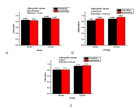
Figure 3: Attenuation of each energy by
a. Bone phantom,
b. Al and
c. Cu samples.
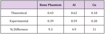
Table 2: Comparison of theoretical and experimental values of mass-thickness.
Conclusion
SrI2 :Eu2+ crystal array coupled with a SiPM array is a viable alternative to CZT detectors used on commercially available DEXA systems for Bone Mineral Density measurements. Our prototype produced comparable or smaller errors between the theoretical and experimental values than results obtained with commercially available systems. Our research will continue focusing on obtaining images of different BMD bone phantoms.
Pre-Competitive Period in Olympic Wrestling Athletes: Losing Weight, a Risk Factor-https://biomedres01.blogspot.com/2021/02/pre-competitive-period-in-olympic.html
More BJSTR Articles : https://biomedres01.blogspot.com


No comments:
Post a Comment
Note: Only a member of this blog may post a comment.