The Impacts of Cigarette Smoking on Rat’s Trachea: A Histologic Study
Introduction
There are many chemicals which are characterized by being
harmful for the human health included in cigarette smoke [1].
Cigarette smoke has been categorized as a human carcinogen that
makes a health threat for smokers and passive smokers [2]. It has
been reported by several studies that the smoke of cigarette as an
aerosol which includes two phases containing gases and particles. In
general terms, cigarette smoke can be considered as a complicated
mixture of oxidants and toxic substances. These studies have
identified 4,800 substances to be included in tobacco smoke among
which are 69 recognized carcinogens as well as a great number of
toxic substances [3]. Smoking is thought to mainly affect lungs and
participate in inducing several diseases such as respiratory diseases
(lung cancer, chronic obstructive pulmonary diseases), cancer of
the breast, brain, stomach, leukemia, lymphomas, coronary heart
and peripheral vascular diseases, inflammation of the arteries,
progressive narrowing of the vascular lumen, risk of developing
myocardial infarction, etc., [3].
The Trachea is a tube that transports air from the upper
respiratory tract to the lower respiratory tract [4]. The cervical
trachea and the thoracic trachea are the two parts of the trachea. The
trachea is made up of 15-20 hyaline cartilage rings [5]. A layer
of pseudostratified columnar epithelium ciliated with goblet cells
lines the trachea. Mucins are produced by goblet cells, which are
unicellular glands that moisturize and protect the airways. Mucus
coats the ciliated cells of the trachea, allowing the cilia to detect
inhaled foreign particles and propel them to the larynx and finally
the pharynx, where they are either ingested or ejected as phlegm.
Mucociliary clearing is the name given to this mechanism. The
ciliated cell has roughly 300 cilia, each with numerous mitochondria
beneath them to supply energy. Brush cells, which have multiple
microvilli connected to their apical surface, are another type of
columnar cell [6].
Objectives
The main objective of the present study is to explore the pathologic changes associated with cigarette smoking on rat’s trachea.
Methodology
Rats were randomly assigned into two groups (n= 8 per group), group 1 was negative control exposed only to fresh air, group 2 exposed to smoking (red LM cigarettes) as 1 cigarette/rat/day for 30 consecutive days. A further period of one-month non-exposure (cessation) to smoking was followed as a recovery stage from the effects of cigarette and waterpipe smoking. Following each period, histological studies were performed. The groups which were exposed to cigarette smoke were divided as follow: Group (2): was exposed to red LM cigarette for one month followed by a recovery period for one month.
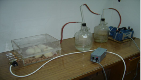
The Digital Smoking Machine
A digital smoking apparatus was designed that have a special
smoking topography, suitable for the exposure of rats to cigarette
smoke [7]. The smoking machine is composed of the following
components as illustrated in (Figure 1). Inhalation chamber made
of Plexiglas (8 mm thick) with the dimensions 30 cm length × 22.5
cm width ×10.5 cm height that can host five rats weighting 100-150
gm.
Each cycle of smoking run lasted for 90 seconds and consisted
of the three following steps:
a) Continuous withdrawing of cigarette smoke for 30
seconds.
b) Washing out of the smoke for 30 seconds, with fresh air.
c) Finally, rats were allowed to breath normal fresh air for 30
seconds.
Following recovery from the last exposure to smoking (overnight) animals were sacrificed by ether inhalation and tissues under investigation were dissected (trachea, lung, and ventricle) and washed with normal saline. Tissues were then fixed in 10% formaldehyde for 24 hours. Tissue was then dehydrated in ascending grades of alcohol, cleared with xylene. Dehydration was achieved by passing tissues through a graded series of alcohol followed by two changes of xylene. After infiltration in paraffin wax, tissues were embedded in pure paraffin wax. Thin sections 5μm thick were obtained by microtome (Spencer 50). Finally, sections were mounted on glass slides and stained with hematoxylin and eosin. Sections were examined and photographed using Zeiss photomicroscope1. Photomicrographs were taken using Moticam 2300 digital camera/3.0Megapixels.
Results
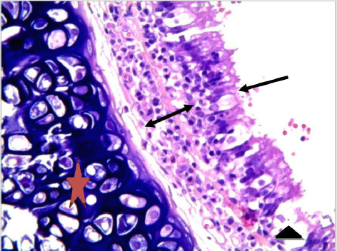
As seen in Figure 2, control sections had shown healthy ciliated pseudostratified columnar epithelium, and all other layers normally seen in tracheal tissue.
Figure 2: Normal tracheal tissue. Cilia (arrow) and goblet cell (triangle). Hyaline cartilage (star). Lamina propria (arrow with two heads). H&E stain. 400X.
Cigarette Smoke-Exposed and After Cessation Period Groups
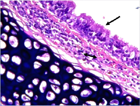
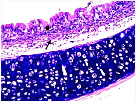
The tracheal mucosa of this group was adversely affected; showing an increase in the number of epithelial cells, amalgamation of cilia, presence of inclusion bodies, heavy lymphocytic infiltration among the epithelial layer was observed and partially induced some structural changes in columnar cells, and goblet cells. Inflammatory conditions were observed through the infiltration of PMNL (Figure 3). After the cessation period. However, lymphocytic infiltration was still present compared to (Figure 3), recovery of cigarette smoking induced some improvements through reducing the level of inflammation and restoring the changes in columnar cells, cilia, and goblet cells with a slight separation in the respiratory epithelium, together with much less amalgamation of cilia were observed (Figure 4)
Figure 3: Tracheal tissue from Rat exposed to cigarette smoke. Infiltration of lamina propria with lymphocytes (arrow with two heads). And partially disrupted cilia (arrow). H&E stain. 400X.
Figure 4: Tracheal tissue of rat after cessation of cigarette smoke. Black arrow indicates infiltration with lymphatic cells. H&E stain. 400X.
Discussion
The exposure to smoking has been associated with adverse mucosal effects in the trachea. These changes included proliferation of epithelial cells, amalgamation of cilia, presence of inclusion bodies, and lymphocytic infiltration within the epithelial layers. structural Changes in columnar cells were also observed as well as in goblet cells. Following smoking cessation most of changes were reversible. These findings are consistent with other studies. In his study, Liao, et al. [8] showed that passive exposure to cigarette smoking in rats induced inflammatory conditions in trachea. The study of Shraideh, et al. [7] reported similar findings in which the exposure of albino rats for 3 months to cigarette smoke induced drastic histological changes in the tracheal epithelium such as epithelial cells proliferation, disruption of cilia, and presence of inclusion bodies.
Conclusion
Cigarette smoking is associated with adverse health effects. Smoking effects were histologically studied on trachea. It was found that in most of changes detected, quitting smoking was essential to revert most changes.
For more Articles on : https://biomedres01.blogspot.com/


No comments:
Post a Comment
Note: Only a member of this blog may post a comment.