Study of the Hypothalamic Pituitary Adrenal Axis Stress Effect on Spiny Projection Neurons by Pathophysiological Computing Modelling of Basement Metabolism, An in Silico Study
Introduction
Medium spiny neurons (MSNs) are a type of inhibitory neuron of the GABAergic type that plays a key role in initiating and controlling body movements so that today these neurons play an important role in the development of neurogenic diseases. Despite the importance of these neurons, there is no accurate information about the effect of stress on them. The subject of this research is a study on the effect of seven stresses include; hypoxia stresses, thermal stress, mTOR stress, oxidative stress, HPA axis stress, aging stress, and electrochemical stress on MSNs by modeling the pathophysiology of these neurons at the system biology level.
Materials and Method
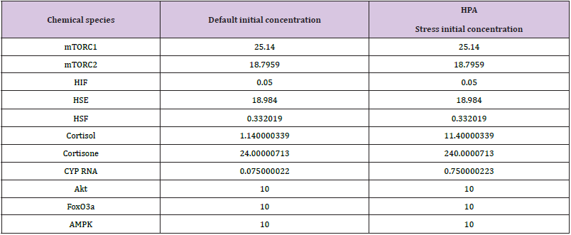
This research can be divided into five stages (Figure 1). In the First step, prefabricated models related to MSN-related stresses, structures, and pathways were reviewed and extracted from biomodel, Vcell, and Reactome databases, and then screened, selected, and archived in SBML format. In the second stage, pathways were merged by COPASI software and a united model was created. In the third stage, the model was implemented on the Vcell platform and the results of the simulation were archived in SBML (level 3, version 1) format. At this stage, the variables were determined to simulate normal and stressed conditions, according to Table 1, and finally, after running the simulator based on solver stiff, the results were plotted by WPS office spreadsheet software and analyzed manually.
Results & Discussion
According to Figure2(C), CYP RNA in stress increased. Cytochrome P450 3A4 (EC 1.14.13.97) is an important enzyme in humans, found mainly in the liver and intestines. This Cytochrome oxidizes small foreign organic molecules (xenobiotics) such as toxins or drugs so that they can be eliminated from the body. While many drugs are inactivated by CYP3A4, some drugs are activated by this enzyme. Some substances, such as grapefruit juice, and some medications interfere with the function of CYP3A4. Use of these drugs with drugs that are modified and enhanced or attenuated by CYP3A4. CYP3A4 is a member of the Cytochrome P450 family. Several other members of this family are also involved in mixed metabolism, but CYP3A4 is the most common [1]. According Figure 2(A,B,F,H,I,P) Dexamethasone metabolites in stress increased. Dexamethasone is a corticosteroid drug. It is used to treat many conditions, including rheumatic problems, some skin conditions, severe allergies, asthma, chronic obstructive pulmonary disease, croup, brain swelling, eye pain following eye surgery, and in combination with antibiotics for tuberculosis.
In adrenergic insufficiency, it should be used with a drug that has more mineral effects (including fludrocortisone). In preterm labor, it may be used to improve outcomes in the baby. It may be taken orally, by injection into a muscle, or intravenously. The effects of Dexamethasone often last for a day and last for about three days. Prolonged use of Dexamethasone may lead to thrush, osteoporosis, cataracts, bruising, and muscle weakness. It should not be consumed while breastfeeding. Dexamethasone has antiinflammatory and immunosuppressive effects [2].
According to Figure 2(G, M, N, O), cortisol metabolites in stress increased. Cortisol is a steroid hormone, a type of glucocorticoid hormone. When used as a medicine, it is known as hydrocortisone. In many animals, it is produced mainly by the adrenal cortex in the adrenal gland [3]. This substance is produced in smaller quantities in other tissues [4]. It is released during the circadian cycle and its release increases in response to stress and low blood glucose concentrations. It also reduces bone formation [5]. This gene encodes the alpha globulin protein with the corticosteroid-binding property. This protein is the major transporter for glucocorticoid hormones in the blood of most vertebrates [6]. PXR (Pregnane X receptor & Glucocorticoid receptor) is a nuclear receptor whose main function is to monitor the presence of foreign toxins and belongs to a family of nuclear receptors whose members are transcription factors that are transcribed by a domain into a ligand and by a Domains are attached to DNA. PXR is a transcription regulator of the Cytochrome P450 CYP3A4 gene.
It is activated by a combination of compounds including Dexamethasone and rifampicin and stimulates CYP3A4 [7,8] The glucocorticoid receptor (GR), also known as NR3C1, is a receptor to cortisol and other glucocorticoid GR is expressed in almost every cell in the body and regulates genes that control growth, metabolism, and the immune response, and because the receptor gene is expressed in different ways, it has different effects in different parts of the body. (Pleutropic) When glucocorticoid hormones bind to GR, the main mechanism of action is to regulate gene transcription [9,10]. After binding to the glucocorticoid, the activated glucocorticoid receptor complex expresses anti-inflammatory proteins in the nucleus. Regulates and suppresses the expression of inflammatory proteins in the cytosol (by preventing the transfer of other transcription factors from the cytosol to the nucleus [11,12]. Steroid Hormone Receptors (SHRs) are transcription factors that are activated in the presence of steroid hormones.
While estrogen receptors are predominantly nuclear, unbound Glucocorticoid (GR) and Androgen (AR) receptors are mostly located in the cytoplasm and are transported to the nucleus only after hormone binding. This Progesterone Receptor (PR ) in humans is encoded by a gene (PGR) on chromosome 11, which has two forms (PRA) and (PRB), that (PRA) is more in the cytoplasm and the form (PRB) in both the cytoplasm and There is in the core. Understanding the mechanism of ATPase activity of HSP90 is largely derived from structural and functional studies of Saccharomyces cerevisiae complexes. Binding of PTGES3 (p23) to the HSP90 complex and, finally, its combination stabilizes the hormone. It is worth noting that GR-importin interactions can be ligand-dependent or independent. In nuclear ligand-activated SHR, specific sequences in DNA called Hormone Responsive Elements (HRE) are created [13]. Albumin is a family of globulin, the most common of which is serum albumin.
All proteins in the albumin family are soluble in water and relatively soluble in concentrated saline. Albumin is usually found in the blood plasma and is not glycosylated. Albumin-containing substances are called albuminoids. Some transfusion proteins are evolutionary linked to the albumin family (including serum albumin, alpha-photo protein, and vitamin D-binding proteins. This family is found only in vertebrates [14-16].
Tyrosine aminotransferase (or tyrosine transaminase) is an enzyme present in the liver that catalyzes the conversion of tyrosine to 4-hydroxyphenylpyruvate [17]. Deficiency of this enzyme in humans can lead to what is known as type II tyrosinemia, in which there is an accumulation of tyrosine (resulting in accumulation of tyrosine due to a lack of aminotransferase reaction) [18].
Conclusion
According to their results and analysis, in general, it can be said that stress caused by the Hypothalamic-pituitary-adrenal axis increases Dexamethasone and cortisol metabolites and increases cellular metabolism and catabolism over anabolism, and therefore can It has a destructive effect on MSNs and other similar cells and thus aggravates the symptoms of MSN-related diseases. It is suggested that researchers investigate various aspects of the destruction of these neurons by researching this subject, and therefore future research could be a follow-up to this research. In this study, we examined only part of the basal metabolism on MSNs, so it is recommended
a) Examine other metabolic pathways not only on MSNs but also on other neurons.
b) Use other bioinformatics software to simulate stress on MSNs and other neurons.
c) Simulation of the effects of different drugs on cell metabolism using relevant software
d) Design of different drugs based on the feedback we receive from the simulator.
Investigation of bio transformation using Biomodel and Vcell.
For more Articles on: https://biomedres01.blogspot.com/


No comments:
Post a Comment
Note: Only a member of this blog may post a comment.