Wednesday, October 12, 2022

Investigating Reliable Recovery Biomarkers of Ischemic Stroke: An Evidence-Based Study

Investigating Reliable Recovery Biomarkers of Ischemic Stroke: An Evidence-Based Study

Introduction

According to the World Health Organization, 15 million people worldwide suffer a stroke each year. Of these, nearly five million die and another five million are left permanently disabled [1]. Millions of stroke survivors are left with very limited motor functions or complete paralysis and depend on assistance [2]. Poststroke survivors majorly suffer from motor, sensory, cognition and language impairments. These impairments occur due to the loss of functions of brain regions surrounding the area of injury due to lesions which further lead to death of neurons of those regions [3]. Majority of cases of stroke are ischemic stroke [4]. Stroke recovery is the complex process due to its heterogeneous nature, where the making choices of treatment and prediction of outcomes and treatment responses are difficult [5].Prediction of outcomes in stroke can be useful to determine which intervention (such as behavioural and therapeutic interventions) will be more effective in stroke patients during the rehabilitation.
However, the methods for accurate prediction of longterm outcomes would allow clinical trials of restorative and rehabilitation interventions to be stratified based on the potential for neurobiological recovery in a way that is not possible if trials are performed in the absence of valid biomarkers [5,6]. A Stroke Recovery Biomarker (SRB) can be defined as an indicator of disease state that can be used as a measure of underlying molecular or cellular processes that may be difficult to measure directly in humans, and could be used to understand the outcome, or predict the recovery or treatment response [5]. Thus, when dealing with a condition as heterogeneous as stroke, validated biomarkers of recovery could help plan treatments and support efficient allocation of resource while maximizing outcome for the patients [7]. Identification of recovery biomarkers shall assist to advance the practice, rehabilitation and recovery after stroke. This systematic review has assessed the role of biomarkers and predictors of recovery in ischemic stroke.

Material and Methods

This systematic review was designed as per the guidelines of Preferred Reporting Items for Systematic reviews and Meta- Analysis (PRISMA) [8-10] and registered with PROSPERO (CRD42020209833) on 18th October 2020 [11].

Eligibility Criteria

Case-control studies reporting the biomarkers and predictors of recovery in ischemic stroke patients in English language published after the year 2000 were included. Studies on animal models, including diagnostic markers, prognostic markers and patients with other psychiatric or neurological conditions (other than stroke) were excluded.

The PICO (Population, Intervention, Comparator, and Outcome) format for this systematic review is:
• P: Patients diagnosed with ischemic stroke and no other psychiatric or neurological condition (other than stroke).
• I: Biomarkers or predictors of recovery in ischemic stroke.
• C: Relative levels of recovery biomarkers in post ischemic stroke patients were compared with the levels of recovery biomarkers found in control subjects.
• O: The most important outcome is the recovery of patients with ischemic stroke. The extent of recovery in ischemic stroke patients is mainly in terms of motor recovery, cognition recovery, sensory and language recovery. Recovery outcomes were measured using the standardized mean difference in level of biomarkers for individual studies.

Information Source

A systematic literature search was conducted using PubMed, Wiley online library, Rehab Data and PEDro Database. First, a comprehensive search was performed individually for all components of PICO. Later individual searches were combined with the help of Boolean operators (AND, OR, NOT). Search was performed with a combination of MeSH (Medical Subject heading terms) and keywords terms. The MeSH terms searched were “stroke”, “Rehabilitation”, “Biomarker” and “Brain Infarction”. Search was performed in September 2020 and was also updated before the submission. Search strategy was developed as per the Cochrane checklist of developing search strategy. [12] Structured strategy for PubMed is given in the Appendix A1.

Study Selection

Studies obtained through initial search from different databases were combined after removing duplicates using the Endnote “find duplicates” filter. A single reader read the title and abstract of all the obtained references. Screening of search records was performed against the predefined inclusion criteria and irrelevant studies were excluded. The reasons for excluding the studies during screening were also documented. Once preliminary articles were identified as potentially eligible, the papers were fully reviewed by two members to determine inclusion; and, disagreements in between were resolved by consensus. In case of multiple publications of the same study, the most recent publication was considered. Bibliographies and citation sections of retrieved articles had been reviewed for additional pertinent studies.

Data Extraction

A pre-specified data collection template was used to extract data from the articles obtained after screening and all disagreements were resolved by discussion among team members. The following information was extracted from the potentially eligible full-text studies;
• Publication details: Author’s name, year, journal’s name, country.
• Population related details: Total enrolled participants, gender, age, days post stroke, stage of stroke, number of participants in case-control groups.
• Intervention related details: Name of biomarker or predictor, type of biomarker or predictor, method to recognize biomarker or predictor, levels of biomarker or predictor in case-control groups.
• Outcome related details: Type of recovery, recovery in casecontrol groups, tools for assessing recovery, National Institute of Health Stroke Scale (NIHSS) score, Modified Ranklin Scale (MRS) score, correlation values in between biomarker/ predictor and recovery.

Quality Assessment

The Newcastle-Ottawa Scale (NOS) was used to assess the quality of studies independently by two authors. The NOS consists of three domains: selection, comparability, and outcome/exposure. It assigns a maximum of four stars for the selection, two stars for comparability and three stars for the exposure category. Thus, nine stars altogether indicate the high quality, seven to eight stars indicate medium quality and six or less stars indicate low quality. Any conflicts were resolved by consensus and individual score of each study was recorded which represents the quality of study.

Risk of Bias in Individual Study

The Cochrane Collaboration’s tool for assessing the risk of bias was used for the risk-bias-assessment at the study level. The assessment was done for the domains namely random sequence generation and allocation concealment for selection bias; incomplete outcome data (attrition bias); selective reporting of outcome (reporting bias); and other biases including publication bias [12].

Data Synthesis and Analysis

The extraction of data from eligible studies was done in Excel spreadsheet, Microsoft Office 2010 (Washington, USA). A meta-analysis of studies was performed through standardized mean difference of recovery biomarker levels. The mean levels of recovery biomarkers or predictors obtained by selected studies were compared in between case and control groups. A value of P<0.05 was interpreted as statistically significant. The I² index was used to assess heterogeneity between studies [13,14]. Random effect and fixed effect models were used to calculate the mean effect size of studies with significant heterogeneity (I² >75%) and without significant heterogeneity (I² <75%), respectively. For systematic review and risk of bias assessment, Review Manager 5.3, Copenhagen: The Nordic Cochrane Centre, The Cochrane Collaboration, 2014 was used [15].

Results

Study Selection

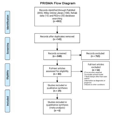
A total of 993 potentially relevant studies were identified using online databases (PubMed, Wiley online library, Rehab Data and PEDro Database) through a systematic search strategy. The 145 duplicates were found and removed after combining the studies obtained from different databases. After screening the titles of 848 studies, 765 studies were excluded, as they were found irrelevant. Further abstract screening of the remaining 83 studies found 32 studies ineligibles as study design was other than case-control, animal models, diagnostic or prognostic markers, based on other conditions and no recovery was assessed. Thus, a total of 51 studies were identified for full text screening. Identified full text studies were retrieved and screened for the eligibility. After detailed evaluation, 29 studies were found to be eligible for final analysis and measuring at least one or more of the considered outcomes in this systematic review [7,16-43]. The PRISMA flow diagram for selection of studies has been represented in (Figure 1).
biomedres-openaccess-journal-bjstr

Figure 1: PRISMA flow diagram representing the selection of studies.

Study Characteristics

The results of this systematic review are based on 29 studies involving 2528 participants. Out of 2528 participants, 926 were in the case group (patients diagnosed with ischemic stroke) and 1602 were in the control group (healthy volunteers). The average age and range of pooled patients were 59.49±5.98 years and 21-93 years, respectively. The results of 12 studies were based on acute stage stroke patients, nine studies on chronic stage, and three studies on sub-acute, two studies on combination of acute and chronic stage and in remaining three studies stage was not specified. Thus, the majority of participants (567/926) in the case group were found with acute stage of Ischemic stroke while, 237/926 participants were with chronic stage. The average months after the occurrence of stroke, when recovery was assessed in participants were found to be 19.36±27.55 months.
The majority of studies (20/29) reported motor recovery while few studies reported cognition recovery (3/29), sensory recovery (2/29), combination of motor and cognition recovery (3/29) and combination of motor and sensory recovery (1/29). Total 27 different measures were used to capture the outcome (recovery) in the participants enrolled in the included studies. The most common used measures of recovery were: General measures- National Institute of Health Stroke Scale (NIHSS), Modified Ranklin Scale (MRS); Motor recovery measures- Fugl Meyer Assessment (FMA), Wolf Motor Function Test (WMFT), Action Research Arm Test (ARAT), Arm Motor Ability Test (AMAT), Motor Assessment Scale, Finger Tapping (FT), Ashworth Scale, Chedoke-McMaster Stroke Assessment (CMSA), Barthel Index (BI), Grip strength, and Edinburg Handedness Inventory (EHI quotient); Cognition recovery measures-Mini–Mental State Examination (MMSE) and Montreal Cognitive Assessment (MCA). The publication, population, intervention and outcome characteristics of all selected studies are presented in the Appendix Table 1.
biomedres-openaccess-journal-bjstr

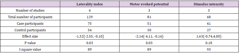
Table 1: Overall effect estimate of laterality index, motor evoked potential and stimulus intensity.

Biomarkers and Predictors

A total of 22 markers were reported in this systematic review. Further, these markers were categorized in sections: Motor Evoked Potential (MEP), Laterality, Cortico-Spinal Tract (CST), brain oscillatory activity, brain connectivity and activation, location, size and volume, cerebral blood flow and others. The details of the markers and their association with the recovery in Ischemic stroke patients are presented in the Appendix Table 2. Predictors like MEP (onset, amplitude, area and selectivity), laterality (laterality changes and index), brain oscillatory activity (event related desynchronization), brain connectivity and activation (resting state functional connectivity, ipsilesional cortico-cerebellar functional connectivity, brain activation and stimulus intensity), CST (damage, weighted CST lesion load) were found positively correlated with the recovery in ischemic stroke. On another hand, predictors like lesion size, infarct volume, lesion topography and white matter integrity were found negatively correlated with the recovery.

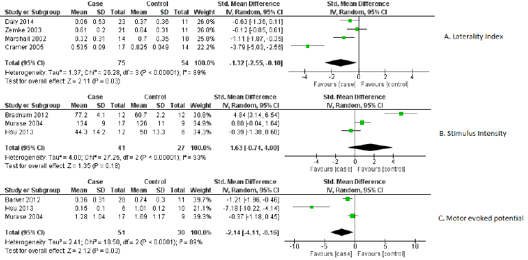
Out of 22 markers, three relevant markers were analysed by performing meta-analysis. The performance of these markers was measured in terms of their capability of differentiating the recovery of ischemic stroke from healthy participants. The standardized mean difference value of markers was used as an outcome measure for differentiation between case-control groups. As a significant heterogeneity was found, therefore the random-effects model was chosen over the fixed effects model. These relevant biomarkers differentiated ischemic stroke’s recovery from controls with good performance: MEP [standardized mean difference -2.14, 95% CI (-4.11, -0.16), P =0.03], laterality index [standardized mean difference -1.32, 95% CI (-2.55, -0.10.), P =0.03] and stimulus intensity [standardized mean difference 1.63, 95% CI (-0.74, 4.00), P =0.18]. The individual study effect sizes were reported in the forest plots based on outcomes. In view of the study wise reporting of markers, sample size was the highest for laterality index (129 participants), whereas the sample size for MEP and stimulus intensity were 81 and 68 participants respectively. The overall effect estimates of laterality, MEP and stimulus intensity were represented in (Table 1). The visual examination through the forest plot reflects that laterality index and MEP markers favours the case group. While the marker stimulus intensity favours the control group. The forest plot of these above mentioned three markers were represented in (Figure 2).
biomedres-openaccess-journal-bjstr

Figure 2: Forest plot of biomarkers
A) Laterality index,
B) Stimulus intensity and
C) Motor evoked potential.

Quality of Studies

Three parameters of quality: selection, comparability, and outcome/exposure were measured through NOS scale. One study was found to have high quality (nine point’s altogether), 23 studies had medium quality (seven-eight points) and five studies were poor quality reports (≤ six points).

Risk of Bias

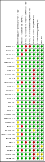
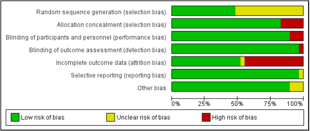
Mainly selection and attrition bias were observed within the selected studies. In few studies reporting and performance biases were also detected as the outcome was found to be reported only for selected groups. The risk of bias was considered adequate for the outcomes. The risk of bias for individual study and their summary were represented in (Figures 3 & 4) respectively.
biomedres-openaccess-journal-bjstr

Figure 3: Risk of bias for individual studies.
biomedres-openaccess-journal-bjstr

Figure 4: Summary of risk of bias.

Discussion

This review has systematically evaluated the biomarkers and predictors of recovery in ischemic stroke as well as their association with the recovery. A number of studies have assessed the association of various markers with the recovery after stroke. Among these, studies with case-control design are growing in number to determine the best predictor which can differentiate recovery in stroke patients from severely affected ones as well as healthy participants. To the best of knowledge, this is the first systematic review based on a case-control design to evaluate the markers which can predict and differentiate the recovery after the stroke. Through the comprehensive literature search, a total of 22 rapidly growing markers were found to be associated with the recovery after stroke. These markers were further categorized into eight broad categories in this review. The results of this review indicate that out of 22 markers, the laterality index and MEP were the only biomarkers that show relevant association with recovery. Hayward, et al. [44] reported MEP as the relevant biomarker to predict motor recovery which is consistent with our results. In addition, the results of this study demonstrated that the laterality index is also strongly associated with the outcome. Single- or repetitive-pulse stimulation of the brain causes the spinal cord and peripheral muscles to produce neuro electric signals known as MEPs [45]. MEPs or their absence serve as indications of the cortico motor pathway’s functional integrity and excitability, facilitating the assessment of associated motor impairment at the time of testing [46]. In this review, different variables of MEP such as MEP onset, MEP amplitude and MEP area were also found to be strongly associated with the recovery. The presence of MEP indicated better recovery and the mean values of MEP amplitude were found to be higher in healthy subjects than ischemic stroke participants.
Aside from MEP, the laterality index was revealed to be a relevant biomarker in this study. The laterality index is a method of determining hemisphere dominance in a range of activities, including language, cognitive skills, and changes in laterality in clinical populations, as in post-stroke condition [47]. In post-stroke fMRI investigations, it has been used to identify neuro plastic alterations in stroke survivors [37]. Further, it was found to be associated with greater activation in the hemisphere contralateral to the working limb (lesioned hemisphere) versus the hemisphere ipsilateral to the working limb (non-lesioned hemisphere) [24]. In this review, the laterality index was mainly extracted from functional MRI (fMRI) in most of the included studies and revealed that stroke is associated with a less lateralized pattern of activation when compared to healthy participants. It further indicated, more the shift towards the normal state of brain function is associated with a better recovery stage after stroke. In contrast to MEP and the laterality index, the pooled values of brain stimulus intensity were identified as statistically insignificant biomarker in this review. Increased stimulus intensity has been linked to a reduction in response time which represents rapid sensory and perceptual processing manifested in the presence of more intense physical stimuli. Further, in stroke survivors, the role of stimulus intensity was also detected in preparing voluntary movements using various techniques such as transcranial direct current stimulation (tDCS) and transcranial magnetic stimulation (TMS) [48]. However, the stimulus intensity for tDCS cannot be individualized based on motor thresholds as for repetitive TMS and may produce variable effects between individuals [39].
In this review the pattern of mean values in the case of brain stimulus intensity was found to be higher in ischemic stroke participants than healthy participants. The mean values of markers of the CST (CST integrity), brain oscillatory activity and connectivity were slightly higher in healthy subjects. The markers like lesion size, location, and infarct volume and lesion topography were not found to have any significant association with the recovery. Authors also want to address the limitations of presented study. Our literature search was constrained to articles only in the English language, which creates the possibility to have somewhat biased results. The outcome measurement in all the included studies was made with a variety of tools and different endpoints. Due to the limited individual patient data, pooling was not possible for all the biomarkers. Thus, it further suggests future studies to investigate recovery biomarkers mainly for cognition and sensory recoveries after stroke.

Conclusion

This review concludes that laterality index, MEP and stimulus intensity are the most relevant biomarkers to predict motor recovery in ischemic stroke patients. The biomarkers of cognition and sensory recoveries, association of markers with individual recovery as well as combination of recoveries (like sensory & motor, cognition & motor and sensory, cognition & motor) need to be investigated more in future studies to predict the recovery with greater precision.

For more Articles on: https://biomedres01.blogspot.com/

Tuesday, October 11, 2022

Comparing the Workplace Organization Method 5s with the 7 Wastes (Muda) in Waste and Failure Management Tool, in the Health Care Quality Management

Comparing the Workplace Organization Method 5s with the 7 Wastes (Muda) in Waste and Failure Management Tool, in the Health Care Quality Management

Introduction

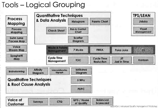
The 5S is a workplace organization system [1] designed to help build a quality work environment, both physically and mentally. The 5S condition of a work area is critical to the morale of employees and the basis of customers first impressions. Management’s attitude regarding employees is often reflected in the 5S condition of the work area. The 5S are: (Redesighningcare, 2017) [1] Seiri (sorting or “SORT”), Seiton (set in order or “STREIGTEN”), Seiso (Systematic cleaning or “SHINE”), Seiketsu (Standardizing or “STANDERDIZE”), Shitsuke (Sustaining or “SUSTAIN”) (Figures 1& 2).

biomedres-openaccess-journal-bjstr

Figure 1: Classification of tools [2].

biomedres-openaccess-journal-bjstr

Figure 2: Before and after implementing 5S Methodology [3].

5S – Step 1 – Sort: Eliminate Items

(Marc Karschies, 2017) “When in doubt, move it out” [2]. Inventories can hold significant values of overstocked, expired and/or useless items. Sort items by logical grouping and get rid of unnecessary or expired items to make space for everything else. If you are not sure about something, put it aside for a few days and see if anyone needs it. Sorting Categories includes: [2]
1. Expired stock (Discard),
2. Replaced by different item/ Never used/ Obsolete items (Discard),
3. Does not belong/ To be returned (Returned to correct dept)
4. Stored elsewhere (Move to correct storage)
5. Overstock to be used (Block reorder until used – Kanban),
6. Mystery items (Investigate and then recategorize),
7. Rarely used (Move to less prominent storage location),
8. Patient specific (Move to special storage) (Figure 3).

biomedres-openaccess-journal-bjstr

Figure 3: (Google images, 2021) [3].

5S – Step 2 – Set in Order, Arrange Items

“A place for everything and everything in its place” [2]. Use containers that hold exactly the right quantity to prevent overstock. Use to bin system – time to restock + buffer = bin size; empty bin = reorder. Label items (with pictures) so everyone can find them or put them away (Figure 4).

biomedres-openaccess-journal-bjstr

Figure 4: (Google images, 2021).

5S – Step 3 – Shine. Neat & Clean

Spot problems quickly by keeping the area spotless [2]. Shine regularly. If something is always in the wrong spot, maybe it needs a new home.

5S – Step 4 – Standardize. Consistent Approach

Make sure everyone is doing things the same way [2]. All drawers organized the same way.

5S – Step 5 – Sustain. Maintain Correct Procedures

When clean in the normal, it is time to see what else you can improve [2]. Perform 5S Audits (e.g with laminated preprinted test scripts like “find XYZ”. Is everything in the right place? Is everything in the right quantity? Perform team huddles to identify further improvement opportunities.

Toyota’s (Ohno’s) Seven Forms of Waste [3]

Taiichi Ohno “father” of the Toyota Production System originally identified seven forms of Muda or waste [3].

Transportation

Every time a product is touched or moved unnecessarily there is a risk that it could be damaged, lost, delayed, etc. as well as being a cost for no added value.

Inventory

Whether in the form of raw materials, work-in-progress (WIP), or finished goods, represents a capital outlay that cannot yet produce an income [3].

Motion

Motion refers to the damage and costs inflicted on what creates the product. This can include wear and tear for equipment, repetitive strain injuries for workers or unnecessary downtime.

Waiting

Whenever the product is not in transportation or being processed, it is waiting (typically in a queue).

Overproduction

Making more of a product than is required results in several forms of waste, typically caused by production in large batches.

Over processing

Doing more to a product than is required by the end-customer results in it taking longer and costing more to produce [3].

Defects

Having to discard or rework a product due to earlier defective work [3] (Figure 5).

biomedres-openaccess-journal-bjstr

Figure 5: (Lean manufacturing tools, 2021).

Methods

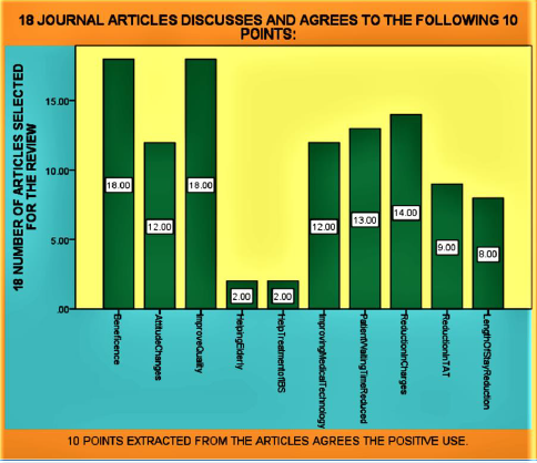
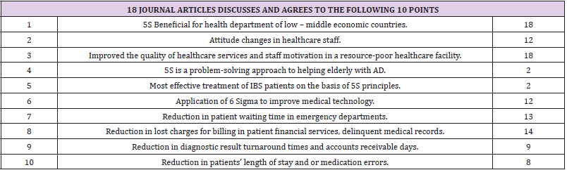
The Author of this article has chosen literature review methodology of 18 random research articles and quality management websites. By reviewing the literature, we have chosen 10 different points in 18 articles discussing or agreeing on where 5S and 7 Muda are beneficial. Most of the chosen articles discusses the quality management in the health sector and their benefits. The 5S management method [4] (where 5S stands for sort, set in order, shine, standardize, and sustain) was originally implemented by manufacturing enterprises in Japan. It was then introduced to the manufacturing sector in the West and eventually applied to the health sector for organizing and standardizing the workplace. 5S has recently received attention as a potential solution for [4] improving government health-care services in low- and middleincome countries. The 5S has the potential to improve [5] client satisfaction at resource-poor health facilities and could therefore be recommended as a strategic option for improving the quality of healthcare service in low- and middle-income countries. To explore more effective intervention modalities, further studies need to address [5] the mechanisms by which 5S leads to attitude changes in healthcare staff.

The pilot intervention of the 5S management method [6] was perceived to have improved the quality of healthcare services and staff motivation in a resource-poor healthcare facility with a disorderly work environment in Senegal. Quantitative and qualitative research based on a larger-scale intervention would be needed to elaborate and validate these findings and to identify the [6] cost-effectiveness of such intervention in low- and middleincome countries. Dental caries and periodontal disease [7] are common in elderly with AD. These dental problems are ambulatory care-sensitive conditions, where effective community dental care can help to prevent the need for hospital admission. 5S is a problem-solving approach to helping elderly with AD [7] build and sustain effective oral hygiene practices for improving their oral health. Irritable Bowel syndrome IBS specialists [8] from around the world established by consensus two best practice charters: the 5S Principles and the 5C Concept. The 5S Principles were conceived to provide health care providers with key guidance for improving clinical practice based on best management approaches. They comprise the following categories: Stage the disease; Stratify patients; Set treatment goals; Select appropriate treatment; and Supervise therapy. Optimized management of patients with IBD based on the 5S Principles can be [8] achieved most effectively within an optimized clinical care environment.

Measure and Statistical Analysis (Table 1 & Figure 6)

biomedres-openaccess-journal-bjstr

Figure 6: SPSS Diagram regarding showing 10 points agrees by 18 articles.

biomedres-openaccess-journal-bjstr

Table 1: 18 Journal Articles Discusses and Agrees to the Following 10 Points.

Results

There is a table and SPSS diagram representing the literature review regarding 18 articles including quality management website. Table title is (18 JOURNAL ARTICLES DISCUSSES AND AGREES TO THE FOLLOWING 10 POINTS). SPSS diagram clearly shows the number of times each article discusses the chosen 10 points. The 10 points includes the agreement of points beneficial for health sector by using 5S and 7 Muda methodology. The healthcare organization is the place [9] where defects and mistakes cannot be tolerated. A simple mistake can cost a human life so defects or mistakes must be eliminated in healthcare service processes. The LSS methodology optimizes the average reduction of a desired process. The expected results can be reductions in several aspects of healthcare such as patient waiting time in emergency departments, lost charges for billing in patient financial services, delinquent medical records, diagnostic result turnaround [9] times, accounts receivable days, patients’ length of stay, or medication errors. Findings lean six sigma LSS is a powerful process improvement [10] methodology that could be applied by health-care sectors to reduce medication errors, increase patient safety and reduce operational costs. Common Lean and Six Sigma tools play a significant role in improving and sustaining the medication process. Practical implications It is necessary for the project team to select the most appropriate LSS tools to address medication process problems. Adoption of a LSS [10] roadmap could help health-care organizations in the successful implementation of LSS. Lean and Six Sigma can be considered [11] valuable process optimization approaches in acute health care settings. The success of their implementation requires significant participation of clinical personnel from the frontline as well as clinical leaders and managers. More research is needed to better understand the factors of success and the barriers to their [11] implementation, as well as their long-term impact.

Discussion

The Medical record department (MRD) [12] is the critical department for the hospital information system and, therefore, the continuous improvement of its services and processes, through scientific methods such as Lean management, are essential. The benefits of Lean for healthcare organizations are that first, the quality of the outcomes in terms of mistakes and errors improves. The second is [12] that the amount of time taken through the whole process significantly improves. Lean and Six Sigma are (Janet H Sanders and Tedd Karr, 2015) continuous improvement methodologies that have garnered international fame for improving manufacturing and service processes. Six Sigma’s Define, Measure, Analyze, Improve, and Control methodology is very similar to good medical practice: first, relevant information is obtained and assembled; second, a careful and thorough diagnosis is completed; third, a treatment is proposed and implemented; and fourth, checks are [13] made to determine if the treatment was effective. The role of Six Sigma philosophy in improvement [14] of the quality of healthcare services is recognized both by researchers and by quality practitioners; discrete-event simulation models are commonly used to improve the key performance measures of patient care delivery. The two approaches are seldom referenced and implemented together however, they [14] could be successfully integrated to carry out quality improvement programs. Six Sigma’s data measurement and process [15] improvement methodology is the impetus for health care organizations to rethink their workflow and reduce malpractice. It involves measuring, recording and reporting data on a regular basis. This enables the administration to monitor workflow continuously. Implementation of the design, measure, analyse, improve and control (DMAIC) improvement cycle, workflow chart, fishbone diagrams and Pareto charts [15] were employed, together with rigorous data collection in the department [16-18].

Conclusion

To conclude the article, 18 of the articles including the quality management websites agrees on the use of 5S and 7 Muda methodology. Different organization around the globe are using 5S and 7 Muda methodology to get benefits for improvement of their health care system. The step-by-step process of 5S and 7 Muda methodology is smart way to start, monitor, finish and follow up the broken health system in several countries.

For more Articles on: https://biomedres01.blogspot.com/

Monday, October 10, 2022

Use of Acetate for the Management of Patients with Staphylococcus Aureus Sepsis

Use of Acetate for the Management of Patients with Staphylococcus Aureus Sepsis

Introduction

Staphylococcus aureus is an opportunistic pathogen that normally colonizes the anterior nostrils of humans, it is one of the most frequent causes of local infections in the skin, where it becomes an entrance to the deeper tissues, where it could give rise to infections fatalities such as sepsis and / or endocarditis [1] Sepsis is the systemic inflammatory response to infection. There are risk factors associated with Staphylococcus aureus sepsis, advanced age, infants, the presence of additional comorbidities such as diabetes, kidney disease, HIV infection, presence of medical devices, application of parenteral drugs Staphylococcus aureus produces clearance from the bloodstream by the immune system, and how this pathogen hijacks the host’s coagulation and defense systems and interacts more with the endothelium of blood vessels where the vessel wall allows the invading pathogen to directly damage the endothelium for its secreted toxins, such as alpha-toxin and superantigens that activate the endothelium, further increasing endothelial dysfunction, aberrant clotting, and vascular leakage [1] The annual incidence of sepsis caused by this pathogen has been increasing in recent years is 4.31 to 38.22 per 100,000 person years in the United States. Their mortality can be 20% despite treatment [2] In the management of fluid resuscitation plays an important role the emergence of the use of crystalloid solutions have predictive benefits and effects on the health of the patient The objective of our work is to analyze acetate management in patients with Staphylococcus aureus septicemia.

Methodology

A detailed bibliographic search of information published in the databases pubmed, Elsevier, scielo, national and international libraries is carried out. The following descriptors were used Staphylococcus aureus, sepsis, acetate. The search for articles was carried out in Spanish and English, it was not limited by year of publication.

Results

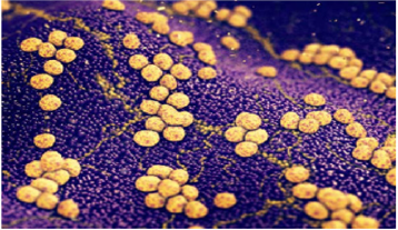
Staphylococcus are a large group of gram positive bacteria, these are distinguished by their groupings that resemble bunches of grapes (Figure 1), this genus of bacteria is characterized by its great adaptability and easy spread [3]. Staphylococcus aureus is part of the normal human flora, between 25 and 50% of the healthy population is colonized by this bacterium, constituting a risk due to its dissemination [4] The pathogenicity of the Staphylococcus aureus species is related to the components of its surface, the pathogenesis caused by the microorganism is due to the decrease in host immunity and virulence factors (that is, the ability to inflict damage to the Guest) [1]. They are the most frequent cause of infections in the skin and soft tissues, the majority are usually selflimited although sometimes it becomes the pathway of entry of the pathogen to deeper tissues and the bloodstream, the presence of Staphylococcus aureus in the bloodstream can lead to sepsis where there is an immunosuppressive response accompanied by inflammation.
In the human body, there is a series of defense mechanisms at the time when staphylococcus aureus enters the bloodstream, it is initially eliminated by the kupffer cells (hepatic macrophages) this process occurs with the help of circulating platelets that they do is bind to the surface of the macrophage, enclosing it for the phagocytization process to be effective. There are strains of Staphylococcus aureus that can evade this process, they can survive and multiply in the macrophage, becoming a source of dissemination [1]. The second line of defense of phagocytosis is activated portal circulation neutrophils, where a small percentage of bacteria can survive intracellularly, thus giving a systemic spread [1]. Later in staphylococcus aureus sepsis, there is an activation of prothrombin and other clotting factors, by the bacteria binding to fibrinogen and using it to cross-link individual cells to form large fibrinogen-coated clumps [1].
Endoletial damage occurs, the microorganism has the ability to adhere to the endothelium, this allows the invading pathogen to directly damage the wall of the vessel by its secreted toxins, such as alpha-toxin and superantigens that activate the endothelium, which further increases dysfunction endothelial, aberrant coagulation and vascular leak [1] Sepsis is one of the most common critical care problems with high morbidity and mortality, it is the main cause of acute kidney injury. Treatment must be essential for the patient to lead to improvement and have less risk and adverse effects [5]. Intravenous fluids are the mainstay of treatment in patients with hypovolemia due to severe sepsis, we currently have colloid and crystalloid solutions for the management of electrolyte therapy. But a controversy has arisen in which we should use, which will bring the best benefit for these patients [5]. Colloids such as dextrans, hydroxyethyl starch, are plasma volume expanders and have been associated with adverse effects such as renal failure with the need for renal replacement techniques, even increasing the risk of death. On the other hand, we found that crystalloid solutions such as Ringer’s Acetate, a clinical benefit with crystalloid is demonstrated in various studies, they contain less concentration of chlorine and sodium [6,7].

biomedres-openaccess-journal-bjstr

Figure 1: Staphylococcus aureus in clusters.

Discussion

It has been shown that part of the action of acetate can occur through the activation of the G-protein-coupled receptor, Gpr43 [8]. These receptors are commonly activated by short-chain fatty acids acetate, propionate and butyrate, ligands that originate from bacteria. anaerobic in the intestine as by-products of fermentation [9], the activation of these receptors allows neutrophils to trigger an antimicrobial response after an additional pro-inflammatory stimulation [10], in addition, the increase in the expression of GPR43 in blood cells is related to a longer survival of septic patients [11]. Through acetate treatment, neutrophils are prepared in a GPR43-dependent manner, leading to increased neutrophil chemotaxis, bacterial death, and better resolution of inflammation and the outcome of sepsis [12]. A study reported that through the action of Gpr43, the apoptosis of neutrophils was induced and the resolution of inflammation, it was also evidenced that acetate regulates acute inflammation by promoting caspase-dependent apoptosis of neutrophils, efferocytosis and leading to a decrease in the inflammatory process inflammation. Resolution of neutrophilic inflammation was associated with decreased NF-κB activity and increased production of anti-inflammatory mediators, including IL- 10, TGF-β, and annexin A1 [8].

Conclusion

Staphylococcus aureus sepsis has a high incidence, mortality, and morbidity rate. Currently there are new drug therapies that can help to have a better evolution of those who suffer from it, where we can highlight the use of sodium acetate; This acts through the Grp43 receptor where it causes apoptosis of neutrophils and therefore the resolution of inflammation. Its use should be more implemented.

For more Articles on: https://biomedres01.blogspot.com/

Friday, October 7, 2022

Successful Treatment of a Chronically Infected and Occluded Aorto-Bifemoral Dacron® Bypass with Bacteriophages

Successful Treatment of a Chronically Infected and Occluded Aorto-Bifemoral Dacron® Bypass with Bacteriophages

Background

In vascular surgery, infections of the vascular grafts are considered to be severe complications [1]. Especially infections of aortic grafts are associated with a high morbidity and mortality of up to 75 % [2]. Since these procedures are often performed in patients with multiple comorbidities, the required explantation of the infected graft and the extensive struggle with the related abdominal infection is related with an early postoperative morbidity and mortality of even over 20 % [3]. Despite the initial achievement of a successful treatment, the general rate of reinfection can be up to 20 % of cases [4]. This is mainly due to bacterial colonies embedded in the peri-prosthetic tissue, which then form a surface-adherent biofilm and hence have an up to 1000-fold greater resistance to antibiotic administration [5]. Even a targeted antibiosis appropriate to antimicrobial susceptibility testing can only suppress a graft infection but does not constitute a curative treatment option [6]. The most common pathogenic bacteria associated with graft inflammation are Staphylococcus aureus, Staphylococcus epidermidis and other coagulase-negative staphylococci, Enterobacterales, Pseudomonas aeruginosa and corynebacteria [7]. These bacteria regularly enhance their specific virulence by attaching to the prosthetic material, and hence averting the local immune response by forming biofilms, that hinder phagocytosis. Furthermore, systemic antibiotic therapy is often inadequate due to the lack of effective saturation concentrations within the inflammatory periprosthetic tissue. In order to reduce the morbidity and mortality associated with the often inevitable surgical treatment, less invasive approaches to adequately treat the infection of the surrounding tissue are urgently needed. In this context, bacteriophages and their bacteriolytic activity are a promising therapeutic option.

Case

In November 2020, a 66 year-old male patient was referred to the emergency ward by his general practitioner with the clinical symptom of an acute abdomen. The examination revealed ubiquitous tenderness on all quadrants with peritonism in the lower abdomen. An infection with SARS-CoV-2 was ruled out. Further examination showed an elevated body temperature of 39.2 °C, and blood testing revealed a leukocyte count of 9.4 x 10⁹/l, as well as an elevated serum C-reactive protein of 90.2 mg/l. The chest X-ray depicted no evidence of pneumonia. An endocarditis was ruled out. Calculated antibiotic therapy with ampicillin/sulbactam was started in the usual dosage intravenously. Blood cultures were positive for Methicillin-susceptible Staphylococcus aureus. Secondary findings included the status of ubiquitous arterial occlusive disease. Due to the necessity of numerous vascular operations on both legs, the patient had eventually undergone a thigh amputation on the right side 12 months earlier; the left side revealed a chronically occluded polytetrafluorethylen (PTFE) Stockmann bypass still in situ.

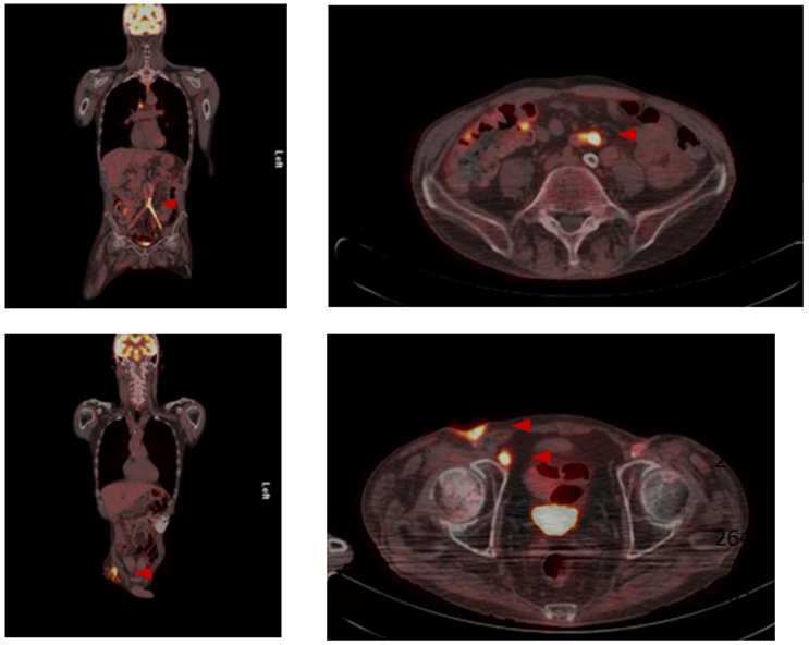
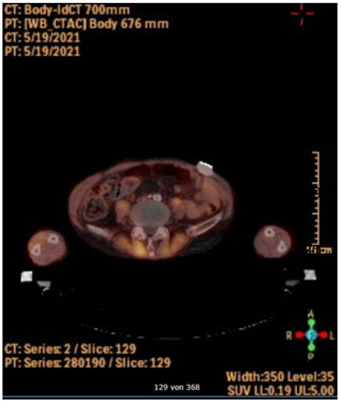
After various transfemoral surgical recanalization attempts in the anamnesis, there were hostile tissue conditions bifemoral with a chronic wound infection leading to exposed graft material. Wound swabs exposed the presence of Staphylococcus aureus and Escherichia coli, indicating a polymicrobial infection. The peripheral blood flow of the lower limbs was compensated. Initially, a CT scan of the abdomen was performed, whereupon an occluded and infected aorto-bifemoral graft was assumed. The subsequently performed PET- CT scan displayed a visibly increased metabolic activity in the area of the graft, so that we diagnosed a chronically occluded and infected aorto-bifemoral prosthetic bypass with subsequential bifemoral infections, leading to the cutaneous wound healing disorder (Figure 1). Due to the patient’s comorbidities, we generally intended an operation and anesthesia time as short as possible with an efficacious treatment by explanting the prosthetic bypass. Further we planned to forego a lavage program for the septic abdomen and intended a primary closure of the abdomen.

In order to treat the local inflammation in the abdominal and femoral areas in the long term intra- and postoperatively, the use of bacteriophages was considered to be plausible alternative therapy option in this case. The patient himself favored an alternative solution compared to an indefinite lasting systemical antibacterial treatment. Therefore, an experimental approach using local bacteriophage application was intended as a last resort treatment in line with Article 37 of the Declaration of Helsinki and in unity with the local ethics committee (A 2021-0208).

biomedres-openaccess-journal-bjstr

Figure 1: Preoperative PET-CT.
Preoperative PET-CT imaging with increased metabolic activity in the area of the aortobifemoral Dacron® bypass as well as the enhancement surrounding the femoral chronic wound infection.

Bacteriophage Treatment

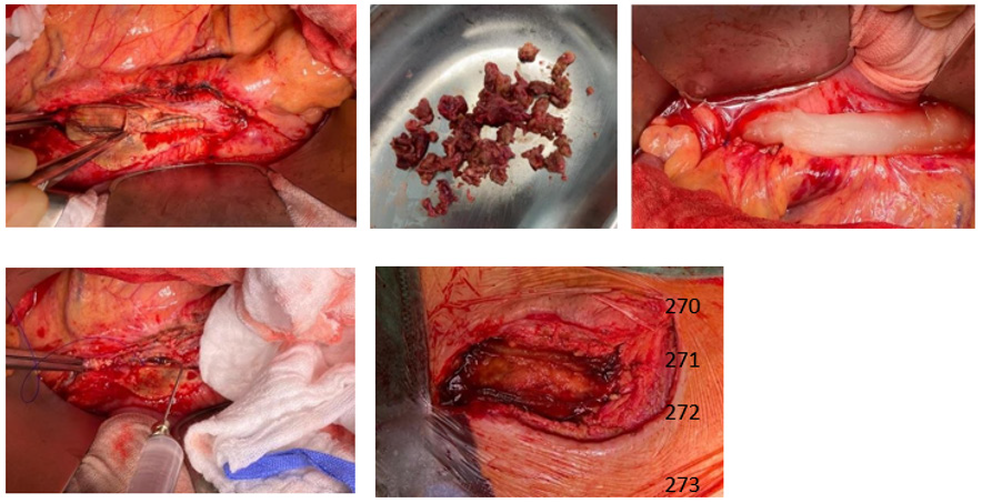
As a curative therapeutic strategy an intra and extra abdominal application of SniPha 360 (Phage24.com, Austria) was executed. SniPha 360 is a commercially available bacteriophage cocktail of lytic bacteriophages against Escherichia coli, Staphylococcus aureus, Pseudomonas aeruginosa, Streptococcus pyogenes, Proteus vulgaris and Proteus mirabilis. After outlining the potential risks but also benefits of the experimental procedure, the patient consented to the therapy. When performing the relaparotomy, cloudy fluid appeared within the abdomen. After an initial lavage, the retroperitoneum was opened, and the proximal aorta was prepared for clamping. The aorto- bifemoral Dacron® prosthesis presented a shell of a biofilm and was embedded in putrid fluid. The infected aortic prosthesis was extirpated, and the aorta was then sutured over. The prosthesis was retrieved femorally after mobilization of the legs of the prosthesis. The bacteriophage suspension was instilled on Tabotamb-Snow®, which was placed retroperitoneally around the infection.

The retroperitoneum and abdomen were primarily closed without further drainage. After removing the femoral anastomoses, the wound conditions were debrided, mobilized and lavaged with a sharp spoon. A bacteriophage-soaked fleece was then placed bilaterally on the femoral side by the same principle, and the wounds were closed again without further drainage (Figure 2). The operation time was 52 minutes, without significant blood loss. Subsequently the patient could be taken to the intensive care unit and extubated without the need for catecholamines. After 10 days of hospitalization, the patient could be discharged with subjective well-being, irritation-free wound conditions and normal findings for inflammatory values in the blood. PET-CT imaging at three months post intervention did not show signs of infection enclosing the aorta or both femoral regions (Figure 3).

biomedres-openaccess-journal-bjstr

Figure 2: Intraoperative images
Intraoperative pictures showing infected aorto-bifemoral Dacron® bypass. Bacteriophage suspension application on a Tabotamb-Snow®, which was placed retroperitoneally.

biomedres-openaccess-journal-bjstr

Figure 3: Postoperative PET-CT
Postoperative PET-CT imaging showing no increased metabolic activity surrounding the bypass.

Discussion

This case demonstrates a successful treatment of a chronically infected occluded aorto-bifemoral Dacron® bypass by a local bacteriophage application. It is assumed that around 50-65% of prosthesis infections are a result of bacterial contamination during surgery [7-10]. A general distinction is made between early (up to 30 days postoperatively) and late infections, although the classification is arbitrary [7,10,11]. Early prosthesis infections are often assumed to be a consequence of intraoperative contamination and late infections to be a result of hematogenous bacterial spread, but profound evidence for this is limited. Late infections are usually caused by insufficient tissue integration of the prosthesis into the graft bed. Common pathogenic agents are staphylococci, enterobacteria and corynebacteria [7,10]. Bacteriophages (or simply ‘phages’; Greek: “bacteria eater”) are viruses that selectively infect bacterial cells and were first described in 1917 by the Canadian Félix Hubert d’Hérelle [12].

Currently Bacteriophages are known as a potent anti-bacterial treatment due to their lytic activity [13]. They are considerably stable when exposed to the inflammatory environment and contribute significantly to the regulation of global bacterial mass. A bacteriophage can only multiply where its host is. They are highly specific and therefore predominantly affect strains within one bacterial species, rarely crossing species boundaries [14]. In the first (lytic) cycle of viral reproduction, phages kill their corresponding bacteria through lysis: once infected, the bacterium host cell then starts the process of reproduction, the destruction of the bacterium, and the release of new phage particles; this process is controlled by enzymes and an interaction of bacterial and phage genes. In the second (lysogenic) cycle, the bacteriophage nucleic acid is integrated into the host bacterium’s genome or forms a circular replicon in the bacterial cytoplasm. Compared to other antibacterial therapeutic strategies like local Rifampine treatment [6], no cytotoxic effects on vascular cells could be found for bacteriophages [15].

In addition, they are effective on multi-drug resistant bacteria as well as biofilm-organized bacteria. Recently, in a case series of eight patients with infections of vascular grafts, surgical wounds or implanted medical devices further demonstrated the feasibility of using different bacteriophages with lytic activity for successful treatment of bacterial infections [16]. Although bacteriophages were used for successful treatment of infections of vascular implants, bacteriophage treatment is still not common and not an officially recommended option for infections in the westernized hemisphere [17]. The retro- and intraabdominal application of phages directly to the infection site ensured a maximum concentration, contact time and invasion of the bacteriophages into the infected peri graft tissue. We were able to perform a short operation time, a definite treatment in respect to complete skin/wound closure and the forego of any drainages. No bacteriophage related clinical adverse events had been detected in our case. A three-month follow-up PETCT scan revealed no signs of infections. It could be assumed that the bacteriophage treatment was successful.

In order to treat the local inflammation in the abdominal and femoral areas in the long term intra- and postoperatively, we perceived the use of bacteriophages as an alternative therapy option in antibacterial local therapy. However, there is an ongoing follow-up for the patient to assure a lasting treatment success. In summary, this case report demonstrates that bacteriophage treatment could be a curative treatment option for patients with bacterial graft- and peri graft infections that are not suitable for extensive surgical approaches.

For more Articles on: https://biomedres01.blogspot.com/

Diagnostic Approach in X-Linked Adrenoleukodystrophy in the Pediatric Patient. A Case Report

Diagnostic Approach in X-Linked Adrenoleukodystrophy in the Pediatric Patient. A Case Report

Introduction

X-linked adrenoleukodystrophy (X-ALD) is the most common peroxisomal disorder, caused by mutations in the ABCD1 locus Xq28 gene due to deficiency of the ALDP protein of the peroxisomal membrane resulting in accumulation of long-chain fatty acids (VLCFA) mainly in the adrenal cortex and central nervous system [1]. It has an incidence of 1 in 21,000 hemizygotes and 1 in 16,800 in heterozygotes. Cerebral infantile X-ALD is the most devastating and progressive phenotype, occurs between three and ten years (peak seven years) is characterized by developmental regression, severe sensory and neurological deficits, in addition to clinical data characteristic of adrenal insufficiency (Addison) [2].

Case Presentation

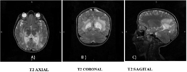
A 7-year-old boy with no major medical or perinatal history. He started his current condition 6 months ago with asthenia and adynamia, loss of previously learned fine motor skills (buttoning pants, tying shoes), decreased interaction with people and impaired academic performance. Likewise, areas of hyperpigmentation were observed in folds (neck, armpits, elbows, English), so he was requested levels of Adrenocorticotropa hormone ACTH levels of 5455pg/ml (normal: 12 - 76pg/ml) and serum cortisol within normal limits for age. Prednisolone was started at physiological doses. He presented an episode of adrenal crisis secondary to community-acquired pneumonia, so the dose was adjusted to stress doses in the previous 5 months. In this hospitalization, contrasted brain Magnetic Resonance Imaging (MRI) was performed, where hyperintensity was found in the T2 sequence at the level of the white matter in the posterior region of the parietal and occipital lobes, as well as at the level of the midbrain and bridge (Figure 1). He was send to the Genetics department with suspicion of X-linked adrenoleukodystrophy. During his follow-up by Pediatric Endocrinology 2 months after diagnosis, he presented progression of neurological deterioration with ataxic gait, impaired verbal communication and urinary and fecal incontinence. The Genetics department who started management with Lorenzo’s oil (40ml/ day) and lovastatin, as well as a diet low in fatty acids, and requested serum determination of very long chain fatty acids (VLCFA) evaluated him. A follow-up was carried out for 3 months, finding poor response to treatment with persistent cognitive impairment, spasticity and rigidity.

biomedres-openaccess-journal-bjstr

Figure 1: Contrast Brain MRI in T2 sequence.
A. Hyper uptake in the region of the internal capsules and parietal lobes;
B. Hyper uptake is observed in the thalamus, as well as in the white matter of the parietal lobes;
C. Hyper uptake is observed in the corpus callosum, in the bridge and mesencephalon.

Discussion

More than 800 variants in the ABCD1 gene mutation are described, which cause defects in ALDP (adrenoleukodystrophy protein) resulting in the elevation of VLCFA mainly hexadecanoic acids (C26:0) and lignoceric acids (C24:0), causing the accumulation of these in the central nervous system among other tissues [1,2]. VLCFA modify phosphatidylcholine in the myelin membrane can cause instability and inflammation due to altered β-oxidation peroxisomal and increased fatty acid elongation, elevated levels of VLCFA in membranes alter myelin structure and function, this mechanism is described in cerebral adrenoleukodystrophy (CALD) [1]. This causes damage to the endothelium of the cerebral microvasculature critical for the initiation and progression of inflammatory demyelination, tight junction proteins have been shown in autopsies to be displaced, along with massive bloodbrain barrier disruption [1,2]. X-ALD is sensitive to oxidative stress that arises from accumulation of VLCFA and when an excess of free radicals occur they cause the opening of the transition pore of mitochondrial permeability resulting in cell death [1]. Clinical phenotypes of ALD have been described: cerebral infantile, adrenomyeloneuropathy (AMN) and primary adrenal insufficiency [2]. Due to the natural evolution of the disease the phenotypes actually represent a spectrum of the condition, practically all males with ALD will develop adrenal insufficiency and progressive myelopathy in adulthood, and may also develop rapidly progressive cerebral demyelination, which can occur during childhood, but also in adulthood [2].

ALD initially presents as adrenal insufficiency between the ages of 3-10 years, of these 35-40% may progress to a rapidly progressive form of inflammatory demyelination (CALD), leading to progressive neurological deterioration with a peak incidence observed between the ages of 3-8 years causing a vegetative state until death within 2-3 years [2,3]. It is clear that other factors modulate this phenotypic conversion in addition that there is an average delay in the diagnosis of adrenal insufficiency of 3.5 years, which is considered a predetermining factor for clinical progression since an asymptomatic period may be followed [2,3]. The presentation of the clinical case had a delay in the diagnosis of more than 6 months, combining the progression of the disease with clinical data such as asthenia and adynamia in addition to hyperpigmentation at the same time demonstrating manifestations of adrenal insufficiency, it is necessary to start the approach by assessing adrenal function [3]. Adding affectation of fine motor skills involvement suggests brain magnetic resonance imaging (MRI) to rule out brain tumor, neuro infection or autoimmune encephalitis [4]. Brain magnetic resonance imaging (MRI) demonstrates demyelination of the brain white matter, which usually precedes clinical symptoms, ALD brain lesions are initially observed in the splenium of the corpus callosum and the parieto occipital white matter [4,5].
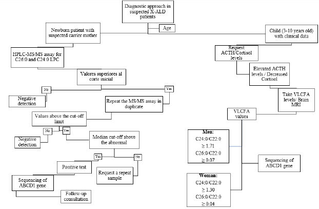
Given the age of the patient, the insidious and progressive onset together with the episode of adrenal crisis provoked by an infection in addition to the data provided by the MRI with which a Loes score of 6 was obtained, it is necessary to approach the patient as a childhood cerebral phenotype of ALD, requesting VLCFA levels [4,5]. Plasma VLCFA analysis is the most commonly used diagnostic test for ALD in men [3]. Given the prevalence of 1 in 21,000 newborns in addition to the difficulty in detecting ADL-X in female patients due to negative family history with onset between the ages of 30- 40 years plus few clinical data (progressive spastic paraparesis more common presentation after age 60) and the need for a panel of laboratory tests [2,3]. In February 2016, newborn screening for X-ALD was added to the recommended uniform screening panel in the United States since ALD has been identified as a newborn screening condition (Figure 2) [2,3]. It was recommended that asymptomatic males identified by newborn screening in New York should have their adrenal function quantitatively assessed every 6 months and the first brain MRI performed until 12 months, then annually until age 3 years, then every 6 months, until age 10 years, and annually thereafter [3]. Once identified, it is suggested to refer the family for counseling, as well as confirmatory tests in the members that require them, in addition, the adrenal function is assessed, since it has been demonstrated that in newborn patients there is biochemical evidence of adrenal insufficiency from 5 weeks and at 4.5 months [2,3].

biomedres-openaccess-journal-bjstr

Figure 2: Proposed algorithm for the diagnosis of ALD-X. It is suggested to investigate clinical history in women with clinical data and perform newborn screening [3]. Normal values VLCFA C24:0/C22:0 0.84μg/mL C26:0/C22:0 0.01μg/mL.

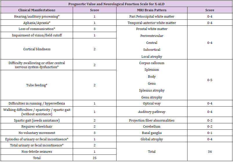
Follow-up with brain MRI is suggested to assess disease progression [3,5]. Abnormalities in MRI are evaluated using the Loes scale, points are awarded based on location, extent of brain parenchymal signal changes, ALD patterns, and the presence of focal and global atrophy [5]. Hyperintensity in T1 or T2 may be seen in areas including white matter: supratentorial, corpus callosum, visual pathway, frontopontine or corticospinal tract and major projection fibers [4,5]. The score is rated from 0 (no evidence of disease) to 34 (severe involvement). 5 In conjunction, the neurological function scale that evaluates the clinical progression of the disease in the patient has been used, in our case resulting in a score of 11 (Table 1) [4]. Both scales should be used to assess progression at a minimum of 1 follow-up evaluation [4,5]. CALD is defined as arrested CALD ≥ 2 consecutive MRI scans within a minimum of 6 months with no increase in Loes Score, no contrast enhancement and no progression of brain symptoms (neurological function scale) [4,5]. There is a limitation in the treatment therapies used in patients with ALD, once the diagnosis is made therapeutic interventions are indicated when the Loes score is between 0.5- 9.2.6 Allogeneic hematopoietic stem cell transplantation is effective for cerebral ALD in the early stages of the disease (Loes 0. 5-9 and neurological function score ≤ 1) allowing to stop the disease progression at the brain level, for symptomatic patients with a Loes score ≥ 10 transplantation produces unfavorable results [4,6]. Transplantation does not alter the progression of adrenal insufficiency so corticosteroid replacement should be initiated, hydrocortisone is the glucocorticoid of choice in children and should be started at stress doses as soon as adrenal insufficiency is diagnosed [3].

biomedres-openaccess-journal-bjstr

Table 1: Assessment of brain involvement. Neurologic Function Scale is used to evaluate the general clinical neurologic status in patients with CALD to determine the degree of risk prior to stem cell transplantation and post-transplant for follow-up [4]. A score of 0 represents no signs of brain disease but more severe signs (*) represent 12 points that determine a high-risk progression domain [4]. Loes < 10 are considered seriously for allogeneic hematopoietic stem cell transplantation at standard risk. Loes > 10 have a more complicated.

Supportive therapies such as Lorenzo’s oil (GTO/GTE) (mixture of glycerol trioleate [GTO] and glycerol trierucate [GTE] in a ratio 4: 1, at doses 2-3ml/kg/day, this reduces the synthesis of very long chain fatty acids (VLCFA) by competitive inhibition of the enzyme responsible for the elongation of saturated fatty acids, but is ineffective in halting disease progression (CALD). Metabolic modulators such as bezafibrate demonstrated a reduction of VLCFA in ALD fibroblasts, but not in plasma, recently a combination of multiple antioxidants at high doses normalizes biomarkers of oxidative damage and inflammation [2,6].

Conclusion

In this case, the importance of making a diagnosis when clinical suspicion persists, recognizing disease patterns with mild to severe patient-dependent manifestations, which are used to assess disease prognosis, as well as the importance of VLCFA analysis to prompt diagnosis of ALD early to allow hormone replacement and prevent disease progression to a devastating phenotype. MRI images of the brain obtained as part of the patient evaluation are used to determine the use of a specific therapy by assessing the risk-benefit ratio, as well as to evaluate progression and determine if the disease is arrested.

For more Articles on: https://biomedres01.blogspot.com/

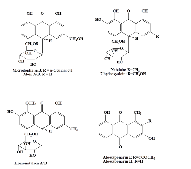
Antimalarial Aloe Compounds

  Antimalarial Aloe Compounds Introduction Among the most prevalent diseases caused by protozoan parasites, malaria is caused by parasites o...