Friday, March 3, 2023

Ultrasonic Frame of Reference for the Downward Displacement of the Posterior and Anterior Leaflets of the Tricuspid Valve in Children


Ultrasonic Frame of Reference for the Downward Displacement of the Posterior and Anterior Leaflets of the Tricuspid Valve in Children

Introduction

In downward displacement of the tricuspid valve [1], displacement of the septal and posterior valves is most common; however, the anterior tricuspid valve can also be moved downward [2]. During ultrasound, the root of the mitral valve in the apical fourchamber view of Ebstein malformation can be used as a reference structure for the downward movement deformity of the tricuspid septal valve [3]: however, there is a lack of reference structures for the downward movement deformity of the posterior and anterior tricuspid valves. Thus, the aim of this study was to evaluate the tricuspid annulus and inferior margin of the coronary sinus were as reference structures for posterior tricuspid valve downward movement. In addition, we aimed to evaluate two-chamber and four-chamber view tricuspid annulus as reference structures for anterior valve downward movement malformation, as well as exploring the reference structure for evaluating the degree of posterior and anterior tricuspid valve downward displacement.

Methods

Selection and Description of Patients

From May 2005 to April 2019, all children with tricuspid valve downward displacement diagnosed by echocardiography in our hospital were selected as study participants. Exclude children with unclear diagnosis. Of them, 18 were male and 24 female patients, aged between 7 months and 15.9 years, with a median age of 3 years and 10 months. Of the 42 total patients selected, 40 also exhibited atrial septal defect. Among them, two cases were further complicated with ventricular septal defect, one with pulmonary valve stenosis, one with ventricular septal defect and pulmonary valve stenosis, and one with single atrium and pulmonary valve stenosis. Two cases were not complicated with any congenital heart disease deformities. These complications are summarized in Table 1.

biomedres-openaccess-journal-bjstr

Table 1: Clinical Information.

Note: ASD: atrial septal defect; VSD: ventricular septal defect; PS: stenosis of pulmonary valve; SA: single atrium

Research Methods

Research Methods

biomedres-openaccess-journal-bjstr

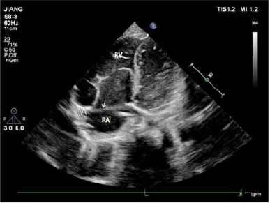
Note: RV right ventricle; RA right atrium

Figure 1: The apical four-chamber view showing that the arrow points to the front of the tricuspid valve annulus. A is the attachment point of the anterior tricuspid valve on the annulus. In the picture, the anterior tricuspid valve is attached to the anterior part of the tricuspid annulus, and the position is normal.

biomedres-openaccess-journal-bjstr

Table 2: Number of patients with different leaflets moving.

Note: SV: Septal valve; PV: Posterior valve; AV: Anterior valve.

Ethical approval was granted by the medical ethical committee of The First Affiliated Hospital of Gannan Medical University with the following reference number: LLSC-202105401. All study participants provided oral informed consent. The tricuspid septal and anterior valves were displayed by echocardiography in the apical four-chamber section, using the ultrasonic examination instrument Philips EPIQ7C and GE VIVID7 (Philips, Amsterdam, Netherlands), with a probe frequency of 3–8 MHz. The descending degree of the tricuspid septal valve was evaluated according to the position of the anterior mitral valve attached to the intracardiac septum, and the size of the atriated right ventricle and right ventricular cavity was observed. The apical four-chamber probe was then rotated about 45° clockwise to make the left atrium, left ventricle, and interventricular septum disappear gradually, exposing the right atrium, right ventricle, and right ventricular posterior wall. The shape, activity and position of the anterior valve of the anterior wall of the right ventricle and the posterior valve of the posterior wall of the right ventricle were observed, the tricuspid annulus and coronary sinus were displayed, and the distance between the attachment point of the posterior tricuspid valve, the inferior edge of the tricuspid annulus, and the inferior edge of the coronary sinus was measured. The apical four-chamber and right cardiac two-chamber views were used to evaluate the downward movement of the anterior tricuspid valve using the tricuspid annulus as the reference structure (Figure 1). The location of tricuspid regurgitation was revealed by color doppler ultrasound (Table 2).

Results

biomedres-openaccess-journal-bjstr

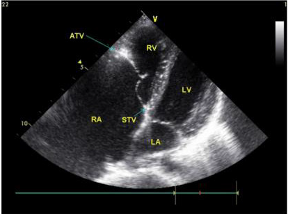
Note: ATV the anterior tricuspid leaflet; STV the septal tricuspid leaflet; RV right ventricle; RA right atrium; LV left ventricle; LA left atrium

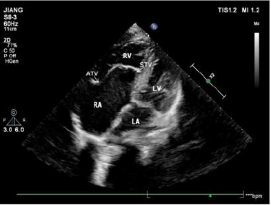
Figure 2: The apical four-chamber view showing that the position of the septal leaflet of the tricuspid valve is significantly lower than the root of the mitral valve during systole.

biomedres-openaccess-journal-bjstr

Note: ATV the anterior tricuspid leaflet; STV the septal tricuspid leaflet; RV right ventricle; RA right atrium; LV left ventricle; LA left atrium.

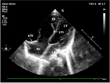
Figure 3: The apical four-chamber view showing that the position of the septal leaflet of tricuspid valve is significantly lower than the root of the mitral valve during diastole.

In 42 patients with Ebstein malformation, the septal and posterior valves moved downward simultaneously in 39 patients; the simple septal valve moved downward in one case; the posterior and anterior valve moved downward at the same time in one case; and the septal, posterior, and anterior valves moved downward simultaneously in one case. Aside from two patients with tricuspid septal and posterior valve downward movement and partial slight downward movement of anterior valve, ultrasound was consistent with the results of operation. Ultrasound showed downward displacement of the tricuspid septal valve in 41 patients (Figures 2 & 3). Aside from two patients in which downward movement of the tricuspid septal valve was so severe that the distance between the tricuspid valve and the root of the mitral valve could not be measured, the attachment point of the tricuspid septal valve was 2.22 ±1.11 cm from the root of the mitral valve in 39 patients.

biomedres-openaccess-journal-bjstr

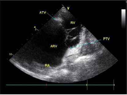
Note: PTV posterior tricuspid leaflet; ATV anterior tricuspid leafle;ARV atrialized right ventricle ;RV right ventricle ;RA right atrium

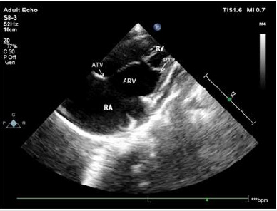
Figure 4: Apical right heart two-chamber view showing that the posterior tricuspid leaflet moves down from the tricuspid annulus and lower edge of the coronary sinus to the apex. The position of the anterior tricuspid leaflet is normal, and the leaflet is elongated.

biomedres-openaccess-journal-bjstr

Note: PTV posterior tricuspid leaflet; ATV anterior tricuspid leaflet; ARV atrialized right ventricle; RV right ventricle; RA right atrium

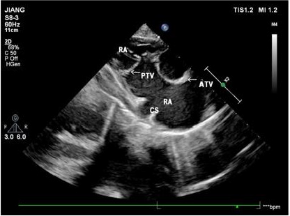
Figure 5: Right ventricular inflow tract view showing that the posterior tricuspid leaflet moves down from lower edge of the coronary sinus to the apex. The position of the anterior tricuspid leaflet is normal, and the leaflet is elongated.

biomedres-openaccess-journal-bjstr

Note: ATV the anterior tricuspid leaflet; STV the septal tricuspid leaflet; RV right ventricle; RA right atrium; LV left ventricle; LA left atrium

Figure 6: The apical four-chamber view showing that the anterior tricuspid leaflet moves down significantly, and the position of the root of the septal tricuspid leaflet is normal.

biomedres-openaccess-journal-bjstr

Note: PTV posterior tricuspid leaflet; ATV anterior tricuspid leaflet; ARV atrialized right ventricle; RV right ventricle; RA right atrium

Figure 7: Apical right heart two-chamber view showing downward motility of the anterior tricuspid leaflet and the posterior tricuspid leaflet positions.

biomedres-openaccess-journal-bjstr

Table 3: Distance of downward movement of different valves.

Note: SV: Septal valve; PV: Posterior valve; AV: Anterior valve.

Ultrasound also showed downward movement of the posterior tricuspid valve in 41 patients with reference to the tricuspid annulus or the inferior edge of the coronary sinus in the view of the inflow tract of the right ventricle and the right ventricle (Figures 4 & 5). Aside from the severe downward movement of the posterior tricuspid valve reaching the cardiac apex that could not be measured in three patients, the distance between the root of the posterior tricuspid valve and the inferior edge of the tricuspid annulus or inferior margin of coronary sinus was 2.71 ±1.08 cm in 38 patients. In one patient, downward movement of the anterior and posterior tricuspid valves, was confirmed by both operation and ultrasound. Ultrasound showed that the position of the anterior tricuspid valve had moved downward in the apical four-chamber section (Figure 6), which was 2.2 cm away from the tricuspid annulus, and that the anterior tricuspid valve was attached to the anterior wall of the right ventricle. The two-chamber view of the apical right heart showed the downward movement of the posterior tricuspid valve (Figure 7), and the distance from the tricuspid annulus was 1.3 cm (Table 3).

biomedres-openaccess-journal-bjstr

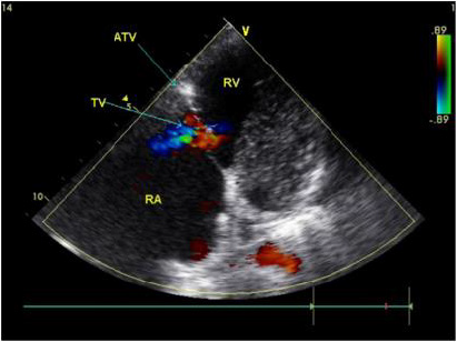
Note: ATV the anterior tricuspid leaflet; STV the septal tricuspid leaflet; RV right ventricle; RA right atrium

Figure 8: Color Doppler showing that the orifice position of the tricuspid regurgitation (blue shunt) has downward direction, and that the direction of the TR flow has an anterolateral bias in patients with downward displacement of the anterior leaflet in the apical four-chamber view.

biomedres-openaccess-journal-bjstr

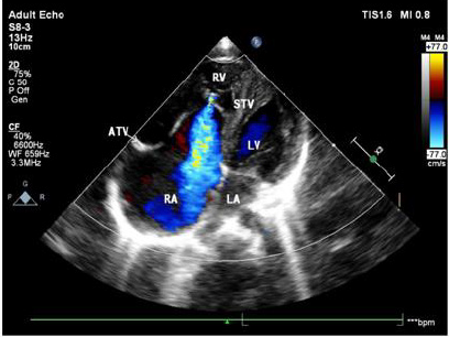
Note: ATV the anterior tricuspid leaflet; STV the septal tricuspid leaflet; RV right ventricle; RA right atrium; LV left ventricle; LA left atrium

Figure 9: Color Doppler in the apical four-chamber view showing that the orifice position of the tricuspid regurgitation (blue shunt) has significant downward motility.

The right atrium was significantly enlarged, the atriated right ventricle was located on the anterolateral side, and the anterior lobe of the tricuspid valve was not obviously lengthened. The apical four-chamber section showed that the tricuspid septal valve was in the exact position. Color Doppler confirmed that the position and direction of the tricuspid regurgitation orifice had shifted to the anterolateral side of the anterior tricuspid valve (Figure 8). Color Doppler ultrasound showed that the position of the tricuspid regurgitation orifice was significantly decreased in 42 patients (Figure 9).

Discussion

In the apical four-chamber ultrasound section, the downward movement of the anterior tricuspid valve shows that the septal valve of the tricuspid valve moves away from the root of the mitral valve, but the reference structure of the posterior and anterior tricuspid valve is rarely reported. All the three valvular lobes of tricuspid valve are attached to tricuspid annulus, while the tricuspid annulus is a cardiac fibrous scaffold structure composed of dense connective tissue. Ultrasound shows hyperechoic light band, and the coronary sinus is located above the tricuspid annulus [4]. The posterior tricuspid valve is distant from the tricuspid annulus in the two-chamber view of the apical right heart [5]. In this study, in 40 children with posterior tricuspid valve displacement, the downward movement of the tricuspid valve was evaluated by ultrasound in the view of the apical right cardiac chamber and the inflow tract of the right ventricle, with the tricuspid annulus and the inferior edge of the coronary sinus as reference structures. Results were confirmed by operation. It is suggested that the downward displacement of the posterior tricuspid valve can be well evaluated by taking the tricuspid annulus and the inferior edge of the coronary sinus as reference structures in the two- chamber view of the right portion of the apical heart.
Downward movement of the anterior tricuspid valve is rare. When the anterior tricuspid valve moves downward, the anterior tricuspid valve is distant from the tricuspid annulus. Ultrasound shows that the tricuspid annulus has a strong echo band, which can show the downward movement of the anterior tricuspid valve. Of the two children with anterior tricuspid valve displacement in this study, one case showed that the anterior tricuspid valve moved away from the tricuspid annulus in the apical four-chamber and two-chamber views of the right heart. The ultrasonic diagnosis of the downward displacement of the anterior tricuspid valve was consistent with the results of the operation. In the other case, the downward movement of the septal and posterior tricuspid valves was diagnosed by ultrasound, and it was found that in addition to the downward movement of the septal and posterior tricuspid valves, there was also a slight downward movement of the anterior valve, which may have resulted from the large area and three-dimensional structure of the anterior tricuspid valve. Among them, part of the slight downward movement of the structure was not related to the change of hemodynamics. The position of the tricuspid annulus attached to the anterior tricuspid valve is sometimes difficult to display. Ultrasound can be expected to show a strong echo light band between the right atrium and the right ventricle from multiple angles, such as the apical four-chamber section, the right cardiac two-chamber section and the right ventricular inflow tract. Detailed attention is required to observe whether the anterior tricuspid valve is attached to the anterior position of the tricuspid annulus. In this study, the ultrasound of one patient with downward displacement of the anterior tricuspid valve showed that there was a hyperechoic light mass in the anterior tricuspid valve attached to the anterior wall of the right ventricle on the apical four-chamber section, which may have been caused by the myocardial echo contrast of the implantation of the root of the anterior tricuspid valve into the anterior wall of the right ventricle. It is suggested that the implantation a of strong echo light mass at the root of the anterior tricuspid valve into the anterior wall of the right ventricle may have been a sign of the downward movement of the anterior tricuspid valve in the apical four-chamber section.
The area of the anterior tricuspid valve was the largest among the three valves, and it was semicircular, accounting for 2/3 of the function. The hemodynamic changes of the downward movement of the anterior tricuspid valve were significantly greater than those of the other two valves, the right atrium was significantly enlarged, the atrial right ventricular wall had become thinner, and cardiac function was poor. Obvious enlargement of the right atrium must sometimes be distinguished from the right atrial aneurysm when the tricuspid annulus and the anterior tricuspid valve move downward at the same time. When the anterior tricuspid valve moved downward in this study, the position of the tricuspid annulus was normal, and only the anterior tricuspid valve moved downwards. The strong echo light mass at the root of the anterior tricuspid valve was implanted into the anterior wall of the right ventricle, and color Doppler showed that the direction of tricuspid regurgitation shifted to the anterolateral direction. This may also have been a sign of downward movement of the anterior tricuspid valve.
The downward movement of the tricuspid valve is more common with downward movement of the tricuspid septal valve, posterior valve, and the lengthy and increased amplitude of the anterior tricuspid valve [6,7]. It is rare that the position of anterior and posterior tricuspid valve is downward, and the position of septal valve is normal, but no matter which valve moves downward, the position of tricuspid annulus remains unchanged, and the position of tricuspid opening remains far away from tricuspid annulus. In this paper, 42 patients with tricuspid regurgitation were shown by color doppler ultrasonography, which suggested that the downward position of tricuspid regurgitation was an important sign in the diagnosis of tricuspid regurgitation.


For more Articles on: https://biomedres01.blogspot.com/

Thursday, March 2, 2023

Cognition and Omega-3 Fatty Acids: A Narrative Review of the Literature


Cognition and Omega-3 Fatty Acids: A Narrative Review of the Literature

Introduction

Today we know quite well that a healthy lifestyle that includes physical activity, healthy eating or moderating toxic habits such as alcohol and tobacco are associated with a lower risk of suffering from some diseases such as cardiovascular diseases, some types of neoplasms, and neurodegenerative diseases in general, and particularly cognitive impairment or dementia. The dietary factor is perhaps the most complex, and much research is aimed at elucidating which foods are associated with this beneficial effect and why. To this respect, one of the most implicated actors in this positive effect has shown to be polyunsaturated fatty acids, especially those belonging to the omega-3 group. Fatty acids are biomolecules consisting of a linear hydrocarbon chain of variable length, with a carboxyl group (-COOH) at one end and a methyl group (-CH3) at the other. The carbon atoms in the chain are joined by single or double covalent bonds. The absence of double bonds defines the acid as saturated, while the presence of one double bond in the chain defines it as monounsaturated acid, and the presence of multiple double bonds as polyunsaturated. Polyunsaturated fatty acids are known by their acronym PUFAs (Poly Unsaturated Fatty Acids). Omega-3 fatty acids (ω-3), together with omega-6 fatty acids (ω-6), make up the group of so-called essential fatty acids, which owe their name to the fact that they are essential for the body since the body is not capable of producing them on its own and must acquire them from foods that contain them. Whether a fatty acid is referred to as omega-3 or omega-6 is established by the location of the first double bond from the methyl-terminal end. In omega-3s, the double bond is at carbon 3 [C3-C4] and can also be identified as n-3. In omega-6, the double bond is at carbon 6 (C6-C7) and is also known as n-6.
Excessive amounts of omega-6 polyunsaturated fatty acids (PUFA), marked by an increased dietary high omega-6/omega-3 ratio, as is increasingly common in current Western diets, are currently speculated to promote the pathogenesis of many diseases, including cardiovascular, cancer, inflammatory and autoimmune diseases [1]. In contrast, omega-3 fatty acids have been shown to play an important role in altering blood lipid profiles and membrane lipid composition and affecting eicosanoid biosynthesis, cell signaling cascades and gene expression, which positively influences health status. This effect seems to have been proven in cardiovascular diseases [atrial fibrillation, atherosclerosis, thrombosis, inflammation, and heart disease, among others], diabetes, cancer, depression, or autoimmune diseases (e.g., rheumatoid arthritis). Its beneficial influence on brain function in the diet of pregnant and lactating women has also been studied [2,3]. The first evidence of this beneficial effect was provided by epidemiological studies which revealed that the traditional Greenlandic diet, rich in marine mammals and fish, reduced the incidence of cardiovascular disease in both the Inuit population and in the Danish people who immigrated to these latitudes, belonging to a different ethnic group [4]. There is currently a tendency in today’s diets to over-consume omega-6 in relation to omega-3 due to the high consumption of vegetable oils by Western society, which means that this ratio can be as high as 20:1, very different from the current recommendations which advise that the omega-6/omega-3 acid ratio should be approximately 4:1, as was the case until the beginning of the 20th century. We could hypothesize that diets rich in omega-3 polyunsaturated fatty acids would be beneficial for the functioning of neural structures, since one out of three fatty acids in the central nervous system are long-chain polyunsaturated fatty acids, and it could be thought that an inadequate balance between these (ω-6/ω-3) would lead to neuropsychological alterations [5].
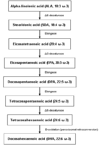
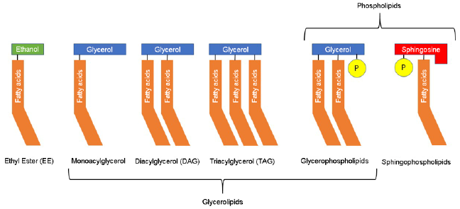
Omega-3 PUFAs originate mainly from the marine environment or the vegetal kingdom and include α-linolenic acid (ALA; 18:3 ω-3), stearidonic acid (SDA; 18: 4 ω-3), eicosapentaenoic acid (EPA; 20:5 ω-3), docosapentaenoic acid (DPA; 22:5 ω-3) and docosahexaenoic acid (DHA; 22:6 ω-3). Some plant seeds, such as flax, chia and canola seeds, are good sources of ALA, which serves as precursor for the synthesis of other long-chain PUFAs (see Figure 1) in the human body such as DHA or EPA. However, the production of longchain ω-3 PUFA from ALA is very limited, with conversion rates of around 5% through this metabolism. Therefore, the best sources of PUFA ω-3 are those of marine origin. Long-chain ω-3s, such as EPA and DHA, are found in the lipids of fatty fish; in the fat tissue of marine mammals; in algae and marine fungi; as well as small crustaceans that are part of krill. The bioavailability of ω-3 PUFAs is also influenced by the organic structure of the ingested form from the diet (see Figure 2), whether ethyl ester (EE), triacylglycerol (TAG) or phospholipids (PL). The most common form of lipids in nature is TAG, which has the best bioavailability compared with EE. PLs are rare in nature and data on their bioavailability are limited and inconclusive [4]. For these reasons, most supplements are made from fish oils or other marine organisms such as krill, as the bioavailability of these products is higher than with vegetal origin products, in addition to the higher proportion of long-chain omega-3 PUFA (DHA or EPA) of higher biological value [6].

biomedres-openaccess-journal-bjstr

Figure 1: Metabolism of ω-3 PUFAs.

The ω-3 PUFAs appear to exert a very important action on neuron membranes, especially in the synaptic regions of neurons (and to a greater extent, in areas of grey matter), where they accumulate in greater proportion and are essential components of the phospholipid membrane, so their importance is vital for the stability of the dynamic structure and functional activity of neurons, as they can alter the fluidity of the lipid membrane (displacing cholesterol from it) and promote synaptic plasticity, which is essential for learning, memory and other cognitive processes. They also act as sources of communication for second messengers between neurons, enhance the coupling of G-proteins involved in many signal transduction pathways and are involved in direct lipid-related transcription functions. DHA, one of the most important and final products of their metabolism (see Figure 1), constitutes more than 90% of the ω-3 and 10% to 20% of the total lipids in the brain. It is mainly incorporated into phosphatidylethanolamine, phosphatidylserine and, in smaller amounts, into phosphatidylcholine in synaptic terminals, mitochondria and endoplasmic reticulum. In fact, DHA is able to modulate cellular properties and physiological processes such as membrane fluidity, neurotransmitter release, gene expression, myelination, neuroinflammation and neuronal growth [6].

biomedres-openaccess-journal-bjstr

Figure 2: Structure of lipids.

The purpose of this review is to examine the current evidence on the relationship between dietary omega-3 supplementation and improved cognition or prevention of cognition-related diseases such as Alzheimer’s disease (AD). To this end, we will discuss the evidence both for and against, in preclinical experiments as well as in clinical studies in humans, including normal ageing, subjective memory or cognitive complaints, mild cognitive impairment, and even AD.

Preclinical Data

The potential effect of omega-3 fatty acids on cognition has been studied in multiple experimental preclinical studies in animals, both in models of normal ageing and in models of AD. The most common model of normal animal ageing is the canine model. Dogs are capable of developing age-related cognitive decline similar to that found in humans and their diet is similar to ours [7]. They can therefore be used to study the potential effects of nutritional supplements in controlled settings. Most studies use nutritional supplements that combine different types of omega-3s, as well as amino acids and antioxidants. The results obtained in these studies support the hypothesis that these supplements would have a beneficial effect on cognition and learning in older animals [7-9], although there are some studies that do not find these benefits [10].
AD models are usually conducted in rodents. These often consist of transgenic strains with mutations that predispose to developing the disease and have been used extensively in AD treatment research. Most studies of omega-3 supplementation in these animal models have shown benefits on delayed cognitive impairment [11], cognitive decline, behavioral symptoms [12], and have even shown to reduce beta-amyloid deposits [12]. An interesting study used transgenic mice for amyloid precursor protein (APP)-animal model for AD-, compared with others that carry the same pathogenic mutation together as well as another mutation that induces the passage of endogenous omega-6 to omega-3, obtaining that the latter showed a lower progression of cognitive and behavioral symptoms compared to the former, displaying the potential protective effect of omega-3 versus omega-6 [13]. This beneficial effect of omega-3s is also observed in other studies in a murine model of epilepsy [14]. In contrast, other studies have not found this beneficial effect in transgenic mice, although they do find it in normal mice [15].

Clinical Evidence

Normal Ageing

We have found and reviewed 13 randomized trials examining the effects of omega-3 dietary supplements in healthy elderly subjects, looking at their cognitive performance. Most used DHA supplements, sometimes combined with EPA, with a daily dose ranging from 350 mg to 3,000 mg of DHA obtained from fish oil in most cases, and in one case from krill [16]. Cognitive performance was measured using a cognitive assessment protocol that typically includes tests of memory, attention, working memory, verbal fluency, and processing speed [17]. The results of most of them showed significant differences when assessing the cognitive effects of omega-3s [16,18-23]. One of the studies used the measurement of P300 evoked potentials as the primary endpoint, resulting in an improvement in the omega-3 treated group [24].
In other studies, on the contrary, no significant differences were found in the parameters evaluated, although most of them used lower daily doses (200-300 mg DHA), compared to the previous studies [25-27], or otherwise the sample size was small [28], which could explain this difference in results. The exception is the study by Danthiir et al. which used high doses (1,720 mg DHA) and only obtained a slight tendency towards improvement in some of the evaluated endpoints [29]. In addition to the above, mention could be made of the Spanish WAHA study, which did not examine the direct effect of omega-3 supplements but studied the effect of a dietary supplement with walnuts in a population-based cohort. Although there were no significant results in cognitive variables after two years of intervention, improvements in functional networks mediated by functional magnetic resonance imaging (fMRI) during working memory tasks were shown [30].

Subjective Memory Complaints

Studies related to subjective cognitive, or memory complaints are scarce, perhaps because the concept is more difficult to categorize or define than normal ageing or the “classic” mild cognitive impairment or dementia. By subjective memory complaints (SMC) we mean individuals who present a subjective perception of poor cognitive performance in general [and memory in particular] but which present a neuropsychological examination within the normal range. It has been established that this altered perception of one’s own cognition could be caused by the onset of a very incipient cognitive impairment which eventually cannot be detected in neuropsychological tests, or by a poor estimation of one’s own abilities (meta-cognition) due to executive dysfunction [31].
The main study in this subgroup of subjects is the MAPT study which randomized French elderly people to multi-domain intervention groups [cognitive stimulation, physical activity, and nutrition], to omega-3 supplementation or both interventions versus placebo. This study found no improvement in patients who underwent intervention [32]. Anyway, a subgroup analysis subsequently found that omega-3 supplementation would be partially beneficial in those subjects who had low baseline omega-3 levels [33]. Another ambitious trial in this population subset is the PONDER study, for which no results have yet been obtained [34]. Otherwise, some studies have found objective improvements on cognitive performance or in regional blood flow measured by fMRI in the posterior cingulum [35-37].
The study by Yurko-Mauro et al. in patients with memory complaints who met criteria for “age-related cognitive decline” has been considered as positive. This is a randomized, doubleblind, placebo-controlled clinical trial in which 900 mg per day of DHA (n=242) or placebo (n=243) was administered to individuals with memory complaints of average age 70 years for a period of 6 months, showing statistically significant differences in favor of DHA administration in validated cognitive tests frequently used for the assessment of memory and learning ability. The authors concluded that DHA supplementation at a dose of 900 mg/day improved memory and learning capacity in individuals with SMC [35]. In addition, there is a meta-analysis evaluating the results of 15 clinical studies, most of them observational, which concluded that DHA/EPA supplementation has a beneficial effect on memory in adult individuals. The review concludes that episodic memory tests of adults with SMC were significantly improved (p<0.004) with DHA/EPA supplementation. Furthermore, and regardless of cognitive status at baseline, DHA/EPA supplementation [at doses >1 g per day] was able to improve episodic memory (p<0.04). Changes in semantic and working memory from baseline were significant with DHA, but no differences between groups were detected [38].

Mild Cognitive Impairment

Mild cognitive impairment [(MCI) is probably the main risk factor for developing dementia, and specifically amnestic MCI (the most prevalent entity) is the most important risk factor for the development of AD. For this reason, preventive treatments have become a relevant source of study in patients with MCI, as they could have a potential role in the prevention of AD. Currently, there is no drug approved for use in MCI, but some strategies such as cognitive stimulation, physical exercise, and dietary recommendations, including omega-3 PUFAs, are under study. Randomized clinical trials that have studied the effect of omega-3 PUFA in patients with MCI have been reviewed. Most studies showed improvements in scores on working memory tests, as well as episodic memory [39- 41], although there are others that evaluated test scores (FSIQ -Full Scale Intelligence Quotient- and WAIS -Wechsler Adult Intelligence Scale), or even depressive symptom scales (GDS -Geriatric Depression Scale) [42,43], which concluded with positive results in favor of omega-3 supplementation. All the mentioned studies above followed patients for 6 months to one year, while studies with shorter follow-up did not seem to obtain statistically significant differences [44].
Additionally, there have been several published meta-analyses and systematic reviews on the effect of omega-3s in patients with MCI. The overall conclusion of all of them points to the beneficial effect of omega-3 PUFA in MCI patients [45,46]. The most recent meta-analysis of 25 studies (n=787) indicated that omega-3s appear to have no effect on overall cognitive function (Hedge’s g= 0.02; 95% confidence interval= -0.12 to 0.154), although it may have a beneficial effect on memory (Hedge’s g=0.31; p=0.003; z=2.945) [46]. Another meta-analysis by Zhang et al. analysed all studies in which the MMSE (Mini Mental State Examination) was used for the assessment of these patients treated with DHA/EPA supplements and concluded that the treatment seems to statistically decrease the rate of cognitive decline in terms of MMSE score (WMD=0.15 (0.05-0.25); p=0.003), so the hypothesis that omega-3 could help to prevent global cognitive decline -in addition to memory- in elderly people with MCI seems to be supported [47].
Neuroimaging research has looked at the effect that omega-3 PUFA may have on the brain in patients with MCI. One of these studies used fMRI to study regional blood flow. It appeared that treatment with omega-3 for 26 weeks is able to increase blood flow in posterior cortical areas, typically affected in MCI [48]. Another study analysed the evolution of brain volume measured by MRI in patients with MCI with results showing that, after treatment with DHA supplementation for one year, the hippocampal volume measured by brain MRI was larger in treated patients than in those receiving placebo [49].

Alzheimer´s Disease

There have been numerous studies on omega-3 PUFA in AD with overall slightly favorable results. In this context, multiple randomized, double-blind, placebo-controlled trials have been conducted, some of which have had great scientific impact. So far, no treatment has been found that is able to significantly improve AD, so most studies are trying to elucidate whether treatments are able to reduce the degree of disease progression. One of the first studies that attempted to address the effect of omega-3s on the progression of AD was the Omega AD study. In this study, patients were treated with high-dose DHA and EPA (1.7 g DHA + 0.6 g EPA) versus placebo, with omega-3 supplementation in both groups continuing for a further period from 6 months to 1 year. The main publication derived from the research showed a tendency for the treatment group to have less disease progression at the cognitive level [measured by MMSE and ADAS-Cog -Alzheimer´s Disease Assessment Scale-Cognitive], although significant differences were only obtained in the mildest AD subgroup [50]. In this trial, a better outcome in patients with higher plasma levels of omega-3 was found [51] and also in those with higher homocysteine levels, which the authors relate to a hypothetical synergistic effect with B vitamins [52]. The same study attempted to assess the efficacy on behavioral symptoms without improvement, with the exception of depressive symptoms, a finding that has been observed in other settings of cognitive impairment [53]. This beneficial effect of omega-3s has been replicated in other clinical trials using similar doses of DHA for 6-12 months with improvements in cognitive scales such as the ADAS-Cog [54–56].
On the other hand, some studies have found no significant differences in terms of cognitive assessment [44]. The most relevant is that of Quinn et al., concluding that treatment with DHA (2 g/day) for 18 months did not produce a relevant cognitive effect as measured by MMSE and subscales of the ADAS-Cog [57]. Studies related to some complex nutrient formulations with high doses of omega-3 PUFA in their composition [1200 mg DHA + 300 mg EPA per 125 ml] are worth mentioning. Some studies with these products have shown beneficial effects of daily treatment. The primary endpoint of the trial was the change in memory as measured by the NTB [Neuropsychological Test Battery] Z-score at 24 weeks of treatment. The study showed statistically significant differences in NTB score in favor of the treated arm [p=0.023; Cohen’s d=0.21; 95% confidence interval (-0.06 - 0.9)) [58,59].

Conclusion

Although evidence is not conclusive, several investigations support that supplementation with omega-3 PUFA, especially DHA, may have beneficial effects on cognition in healthy older adults with SMC and even in patients with MCI or AD. These effects should generally appear at high daily doses [800-900 mg and above] and over long periods of time (6 months or longer). This protective effect has not been shown in all studies, so it is possible that the results may be influenced by other variables; in this sense, it is important to mention that ensuring adequate long-term adherence to treatment [at least 6-12 months] may be important to establish a possible objective benefit. Anyway, more soundly designed interventional clinical trials are needed to ascertain relevant issues concerning dose and supplementation duration. It has been found that plasma omega-3 values or homocysteine levels may be related with these discrepancies among different studies, with subjects with lower plasma omega-3 and homocysteine levels benefiting more from this effect. In any case, these supplements appear to have no relevant adverse effects and may have other added benefits such as reduced cardiovascular risk. Moreover, their indication is not at odds with other recommendations such as cognitive stimulation and physical exercise, so that, altogether, these measures can be medically recommended.


For more Articles on: https://biomedres01.blogspot.com/

Wednesday, March 1, 2023

A Review on the End-of-Life Care


A Review on the End-of-Life Care

Background to the Study

There is a huge distinction between death occurrences and believes in the past and that of the present. The level of death occurrence in the present is increasing compared to the past, and it is influenced by population increase, globalization, global warming, internet penetration, and various discoveries. According to Carlet et al (2004), death is frequently a private, religious or spiritual event with a group of family members and friends gathered around the victim. However, as death becomes a topic of public concern and more technical procedures are introduced to avoid or facilitate dying, the picture of death is changing [1]. End-of-life care for families and their patients has been a hot topic for the past two to three decades. This could be in consequence of a variety of factors, such as the shifting away from a solely medical perspective and toward a more holistic approach to caring. [2], for example, said that the early 1970s saw a transition away from a strictly biomedical survival perspective and toward a dual perspective. This includes focusing care on saving lives and enhancing medical identities, as well as taking care of family members and developing numerous social identities. Supporting life procedures and modern technology introduced to the treatment process contribute to hospitalized patients’ disease becoming more severe [3].
Most significantly, in today’s healthcare environment, families and their patients expect to be included in the treatment process as well as in the decision-making process [4]. The majority of the literature fails to define end-of-life or end-of-life care precisely. However, several authors have attempted to provide a succinct definition of this phrase. These two descriptions appear to be general and ambiguous. It seems to be difficult to establish a precise or comprehensive description of end-of-life care. End-of-life care in the intensive care unit (ICU) was detailed in detail by Alhalaiqa, et al. [5]. Alhalaiqa, et al. [5] pointed out that while delivering endof- life care, there are no set processes or stages to follow, making it difficult to assess the effectiveness of any end-of-life programs. However, talking about death and end-of-life care, as well as having open dialogues about these topics, may contribute to the development of guidelines for healthcare professionals to provide high-quality end-of-life care [6]. End-of-life care is also implemented differently depending on a variety of criteria, including the patients’ conditions and the departments. Cardiopulmonary resuscitation of victims and their relatives in acute settings is the focus of this study.

Literature Review

Stages of the Crisis at a Duration of Unexpected Death

There are four stages of crisis during a sudden death. They are recognized and listed in order of difficulty from the least challenging to the most challenging crisis. If any of these stages are not addressed, a person may progress from one to a more challenging one.
First stage: The presence of a threat that could harm patients’ family members is included in the first stage. This threat may cause stress or tension among the relatives. This stage may entail family members’ fears that something dangerous has occurred to their loved one in the case of cardiopulmonary resuscitation (CPR). The routine coping mechanism, according to Wright (1996), may be able to settle this stage. Wright also stressed the necessity of previous experience in minimizing the effect of this stage. Surprisingly, many studies revealed that most of family members who have previously watched family-witnessed resuscitation (FWR) wish to be present again.
Second Stage: Failure to treat persons in this stage may result in their illness worsening and sending them to the second stage, according to Wright (1996). The failure of previous experience to tell people how to deal with this new situation may have resulted in the second stage. Feelings of helplessness and hopelessness may intensify throughout this stage. People at this stage, according to Wright [7,8], try to find alternatives. Wright revealed that relatives can utilize the trial-and-error method to figure out the most acceptable response to the situation. Allowing FWR was supposed to increase death acceptance and assist bereaved families [9]. This would hasten the grief process and make it easier to accept death as a natural result of CPR.
Third Stage: Intense activity, disorder, and disarray characterize the third stage; old fears resurface, preoccupation with trivia, and make-or-break action are all present. This stage, according to Wright, might be overcome by changing the focus. Wagner. Several studies have found that family members prefer to attend their loved one’s CPR because it allows them to acquire information more rapidly. This would also lessen family tensions and reduce the likelihood of inappropriate behavior.
Fourth Stage: The patient’s relatives may be unable to cope with the situation in the fourth stage, and they may retreat and feel gloomy. Wright recommended taking various actions to manage this stage, including addressing the relatives’ comfort requirements, listening to them without passing judgment, creating a quiet environment, and taking a break from the problem-solving effort.
These interventions were suggested in the literature as a way to satisfy the requirements of the patients’ relatives during CPR. The necessity of treating families as soon as possible would be highlighted if healthcare providers considered these stages and recognized each one. This prevents families from progressing to a more mature level. Individuals do not, however, necessarily progress through all of these stages. Some people may skip the early stages and proceed straight to the advanced stages, while others may adjust to abrupt death and recover swiftly without experiencing any of the preceding stages. The determinants of grief and the elements that affect bereaved individuals during sudden death are discussed in the next section.

Causal Factors of Grieving

In general, there was no systematic study of the elements that would affect families’ reactions and feelings during and after CPR, especially if the CPR resulted in death, in the literature. It might be worthwhile to go over these points again. Several elements were thought to play a role in creating the powerful feelings, reactions, and grief experienced by people who died suddenly. Six elements, according to Wright (1996), have a significant part in deciding how families grieve when a cherished person dies suddenly. These factors include:

a. Manner of Death

The patient’s condition at the time of death should be known to healthcare personnel. It was generally assumed that natural (normal) death would cause less grief than unnatural (abnormal) death. The natural deaths are usually unavoidable, while the unnatural deaths are usually avoidable. The avoidable deaths are mostly caused by either human or mechanical faults. Parkes, who worked as a psychiatrist, noted that all of the bereaved people he had visited had experienced extremely traumatic kinds of bereavement. It has been stated that healthcare professionals who deal with CPR in consequence of injuries and accidents are more rigorous about allowing family members to participate in CPR. The majority of these health care providers raised serious worries about the presentation’s potential adverse effects on family members.
Helmer, et al. (2000) revealed that comparing trauma patients to those with medical disorders is problematic. It would be more difficult to blame others if the death was caused by natural causes such as disease, according to Wright (1996). Family members, on the other hand, may blame healthcare experts, health organizations, culture, or society, as well as themselves for not being able to do more than they did. The sadness of losing a loved one will be magnified if he or she was far away from home or family members, according to Wright (1996). This demonstrates the importance of family members being present during CPR.

b. The Identity of that Person (victim)

The depth of the grieving process is influenced by the patient’s status in the family [10]. Wright (1996), on the other hand, cautioned against making assumptions based on this characteristic. For instance, if a breadwinner dies unexpectedly, such person will be mourned for a long time because of the significant position that the person occupied in the family. Furthermore, the loss of one of the parents would have an impact on other susceptible family members’ feelings of stability and safety.

c. Manner of Attachment

The loss of a key individual is usually the most distressing. This can involve the death of someone with whom one has had a close relationship. As a result, the stronger the link between the grieving individual and the deceased person, the more severe the adverse effects on the departed relatives are expected. This, however, is not a rule. A father or a mother, for example, may have various amounts of attachment to their sons. To determine the strength of this relationship, a series of questions should be asked. This includes questions like, “What does this loss represent?” and “What security or safety aspects might be jeopardized in consequence of this loss?” Wright (1996) noted that the provision of answers to the following questions would aid in determining the bereaved person’s level of vulnerability. Another essential aspect of the attachment’s nature is the security and safety difficulties that arise in consequence of the relationship (Wright, 1996). For example, losing a father who is the family’s sole source of money might increase tension among family members.

d. Past Precursor

Wright (1996) emphasizes the importance of previous crisis experience, particularly abrupt death, in coping with the new crisis. When people have a positive experience, Wright says, it helps them cope with the new incident, and vice versa. To put it another way, what one person considers a loss may not be considered a loss by another. It’s possible that being exposed to the crisis will help people build skills and experience in dealing with similar situations in the future. It was discovered that family members with prior FWR experience were more confident in their attitudes during CPR. Furthermore, those who had been exposed to a comparable situation or had died suddenly were more cooperative with medical personnel performing CPR. They also provide assistance and comfort to the other families.

e. Individual Characteristics

Personal qualities have a vital influence in determining how people react when they are dying or receiving CPR. People react to abrupt death in different ways, according to Wright (1996). Wright also discovered that the personality of a patient’s relatives has an impact on how they search for a healthy grieving resolution. Emotional and physical well-being, according to Walsh and Crumbie (2007), provides people with more resources to cope with losses. As a result, healthcare providers should be prepared to cope with people who have a wide range of mental and physical resources for coping with loss, as well as diverse levels of social support. It appears that assessing people’s personalities or evaluating the differences between patients’ families is challenging during CPR. It appears critical to emphasize that not only personal traits influence people’s reactions, but also other elements such as family preparation, healthcare preparation, and environmental preparation. Family members should, for example, be accompanied by trained staff during CPR [11], and they should be given a suitable location to sit and observe the treatment [11].

f. Social, Cultural and Religious Factors

When people lose a loved one, social, cultural, and religious variables may have a big impact on their feelings and reactions [12]. The availability of social assistance would help to mitigate the crises’ impact on patients’ families. In a survey done by Al-hassan and Hweidi (2004), relatives of Jordanian patients rated their need for assistance as the lowest. Jordanians, it was revealed, rely on other family and friends for the majority of their support. Jordanian people in critical care units were assessed in this study, which was conducted in Jordan. However, the goal of this study was to look at the needs of critically sick patients’ families, not their needs at the time of death.
Some religions and cultures, according to Wright (1996), assist the bereaved by providing support and confirmation of their worth. Some religions, such as Christianity, are commonly thought to be helpful and supportive in the event of a sudden death, according to Wright. Jordanians’ opinions and behavior on matters such as health and death are heavily influenced by religion and culture. People in Jordan, for example, rely on their relatives for financial and psychological support rather than relying on other resources such as hospital professionals. As a result, Jordanian patients frequently have a significant number of visitors. The task of healthcare professionals is made more difficult by the fact that they must deal with a huge number of visitors.
In conclusion, these six criteria appear to be critical to consider when devising any remedy to lessen the harshness of the grieving process. Understanding these characteristics will aid healthcare providers in identifying the issues that patients may need to address in order to begin the grieving process. The following discussion looks at a suitable plan for treating bereaved family members appropriately.

Dealing with Bereaved Relatives at the Time of Sudden Death and During CPR

In the work of literatures, there is no mention of FWR in relation to end-of-life care. According to Kubler-Ross, grievers go through five stages (1969). Denial or a sense of isolation may be felt by grievers at first. This means that grievers may say things like “don’t say that” or “no, he didn’t die” to indicate their disbelief. Wright (1996) suggested a number of methods for coping with this emotion, including finding a polite way to inform family members of the bad news. According to Davidhizar and Newman-Eiger, nurses, on the other hand, should comprehend the value of denial (1998). They claimed that denial is one of the safest strategies to deal with the unfathomable.
Second, grievers may have a sense of rage. They might start saying things like ‘why me?’ or ‘why now?’ as a form of protest. Kubler-Ross warned that grievers might start blaming the healthcare experts for the lack of justice. Nurses and other professionals may take this personally (Wright, 1996). Bereaved people may begin haggling with healthcare providers. They might try to put off the inevitable. Healthcare practitioners, according to Wright (1996), should accept this and endeavor to reach an agreement with grievers to prove the death. Bereaved people may experience depression. When grievers can no longer deny or relocate, they enter this stage.
Grievers may experience feelings of sadness and crying at this stage as they begin to recognize reality. Withdrawal, stillness, and helplessness may be observed by bereaved people. As grievers reach the acceptance stage, they stop striving to ignore or avert the unavoidable death. Grievers begin to comprehend the concept of death at this point, and they begin to relax and feel at ease. Research by Brysiewicz, et al. [13] employed a semi-structured interviews to examine the ED healthcare personnel’ capability to handle the situation of sudden death. The study reacted to findings of previous studies that was conducted by Brysiewicz [14]. A model was created to give healthcare personnel guidance on how to cope with unexpectedly bereaved families before, during, and after death. This approach instructs healthcare providers on how to cope with family members while performing CPR. This would also make it easier for families to accept their loved one’s death. Before the happiness of death, the first half of this paradigm includes instructions for dealing with bereaved families. This comprises implementing two ways to improve the department’s performance, as well as enhancing the department’s culture and guaranteeing enough resources. The model’s second section contains suggestions for coping with bereaved relatives after they have died. This involves making the caring process more efficient. Three ways are expected to do this. Proximity, sensitive communication, and sensitive deathtelling are the three.
The model’s final piece includes advice for dealing with bereaved families when a loved one has passed away. This entails giving family members the best possible support. It was recommended that two approaches be taken. Assisting and supporting medical professionals, as well as assisting mourning families, are among these responsibilities. Once this paradigm was established, ED professionals’ capacity to communicate with families at the time of a loved one’s death was stated to improve [13]. Data from a prior study was used to develop this model. The use of qualitative design allows researchers to gain valuable insight into the perspectives and recommendations of families and healthcare providers. However, because of the small size of the original study’s sample and the fact that the data was taken from a single institution, the findings are limited in their generalizability. This model was implemented in ED. The current study, on the other hand, adopts a different approach. Regarding the effects as influenced by technology, the majority of studies and reviews focused on FWR in emergency departments, according to a review of the literature. The current study, on the other hand, is concerned with the views of healthcare workers and family members in adult critical care settings. As a result, it appears that some light should be shed on nature and the characteristics of the critical care environment. In terms of the rate of CPR, patient conditions, and work environment, Demir [15] highlighted that there are certain distinctions between the ED and critical care units. Critical care units (CCUs) are specialized units for patients with lifethreatening illnesses [16]. During the care of a critically ill patient, healthcare workers are expected to face numerous physiological and psychological problems.
Furthermore, critical care specialists are increasingly expected to provide psychological and emotional support to the families of critically sick patients [17]. Critical care workers, according to Offord [18], are expected to deal with dying patients and bereaved relatives more than experts in other departments [19]. The conditions of patients in critical care units differ from those in other departments. In critical care settings, CPR is a regular technique. Hadders [20] found that ICU clinicians are frequently unsure regarding the resuscitation results of their patients. According to Hadders, individuals who receive CPR either survive or recover completely. After CPR, most survivors rely on machines and technology to keep them alive. According to Benner, et al. [17], the necessity of providing psychological and emotional care for patients and their relatives in critical care settings is undervalued. More than anything else, this was intended to result from a focus on the patient’s biological demands. It should be noted, however, that the presence of a family member in one of the critical care units will upset established family roles and will frequently throw a family into disarray [21].
Moving forward, a number of studies have demonstrated the necessity of assisting critically ill patients’ families and include them in patient care [22]. The critical care environment differs from other hospital departments in that it typically contains skilled healthcare staff as well as advanced technology [16]. In these situations, multiple machines and monitors must be present surrounding each patient. Professionals encounter a number of obstacles in this context. Medical experts had to learn how to use all of the new machines and technologies to begin with [16]. Dealing with technology should not prevent healthcare workers from considering other patients’ and family members’ psychological needs [16]. Sundin-Huard (2005) noted that critical care personnel are frequently obsessed with their patients’ immediate physical and technological needs [23]. To ensure patient survival, technology should be employed to provide maximal patient benefit while also considering the needs of other patients and their families [16].
Mosenthal, et al. [10] noted that people are increasingly seeking death dignity without unnecessary using life-prolonging gadgets, but they equally value high technology’s promise of cure and spectacular lifesaving measures. As a result, it appears that providing technical assistance to critically sick patients and their families, as well as describing the role of each machine in the patient’s environment is enhanced. In a critical care setting, Hadders [20] explained how critically ill patients and their families felt about being reliant on technology. FWR has been reported to be more acceptable in the ED than in critical care settings in the literature. According to Bennun [16], critical care specialists place a greater emphasis on technology than on providing psychosocial treatment to patients and their families.
Because critical care specialists are so focused on technology, they overlook other parts of care, such as family-centered care [24]. This was also assumed to be the reason why critical care workers resisted allowing family members to participate in treatments like CPR [25]. The current investigation takes place in a critical care setting. The majority of the literature, however, is based on evidence from ED settings. As a result, it appears that the findings of these investigations must be taken into account. However, the mind should be awakened to consider the contrasts between the emergency department and critical care settings.

Empirical Review

Mcmahon-Parkes et al. (2009) examined the opinions of patients who survived CPR and those who were not resuscitated. Patients were indifferent about compromising confidentiality in consequence of FWR, according to the researchers. Redley, et al. [26] noted that the ethical principles surrounding FWR should be further discussed. The importance of a qualitative approach in studying FWR would provide a broader perspective on these principles and their impact on healthcare professionals’ and families’ perceptions. Several questions must be answered, such as “who is the person that will be authorized to witness CPR?’ How many people should be present while CPR is performed?’ How about the other family members? Who will look after them? Would additional relatives be willing to stay outside the resuscitation room with you?” All of these questions should be addressed with consideration for the culture of the responders.
A qualitative approach would reveal more information about what family members wish to accomplish while in the resuscitation chamber. Allowing FWR, for example, could affect the public’s trust in the medical profession, according to Rosenczweig [27]. However, the manner in which this would occur was not specified. Other topics are expected to be examined more if a qualitative approach is used. Fulbrook, et al. [11] stressed the importance of recognizing the differences in healthcare systems between countries, which can have an impact on outcomes. In addition, it was identified that the role of self-assurance in accepting or rejecting FWR be investigated [28-30]. They found that nurses’ opinions toward FWR are unaffected by previous experience with the procedure. This finding differs from that of other studies, which found a link between a lack of experience with FWR and unfavorable or doubtful attitudes [31,32]. Fulbrook, et al. [11] advocated for more research into the aspects that influence how people make decisions about the FWR. Many studies about FWR were evaluated by Redley, et al. [26]. They claimed that FWR might infringe on a patient’s privacy. Because there is a scarcity of information about ethical principles from the perspective of patients, these principles may be questioned.
Ardley, et al. [9,33-36] discovered that the majority of the research used quantitative designs. These findings are consistent with other studies that analyzed numerous empirical investigations on FWR. However, it is agreed that this is insufficient rationale for choosing a qualitative approach. As a result, other flaws in using a quantitative technique to examine FWR should be identified. In general, quantitative research is thought to be reductionist [37]. This means that using a quantitative approach will leave some variables unaddressed. As a result, using a qualitative method should provide a more holistic view of the subject under inquiry [38,39]. Fulbrook, et al. [11] investigated the opinions of European Nurses that are saddled with the responsibilities of critical care concerning FWR. Furthermore, some researchers acknowledged the influence of culture and religion on people’s opinions toward FWR [40-42]. These topics, in contrast require further investigation and debate. To do this, healthcare professionals and the general public must be encouraged to engage in open debate and free discussion on FWR [43,44]. Most of the above problems could be explained by using a qualitative design.

Specific Literature Review on FWR and Research Gaps

In the study of Axelsson, et al. [45], six studies were undertaken in the United States, four in the United Kingdom, one in Australia, and one in Sweden. Alhalaiqaa, et al. [46] identified that only two studies were conducted from a Western point of view, and that all these studies may not have considered the views of emergency situation professionals. Also, none of them were conducted in Africa, and none of them supported FWR. However, cultural differences are expected to emerge inside Western countries as well [47]. Fulbrook, et al. [11] examined the attitudes of critical care nurses concerning FWR in Europe. In their studies, they noticed that there were certain differences between British nurses and nurses from other European countries. Walker [36] noted that the global movement to study FWR should focus on cultural differences not only between nations, but also inside the local and national healthcare systems.
Davidson, et al. [23] also noted that the impact of spiritual and religious beliefs on patients’ healthcare decisions has not been extensively investigated in the works of literature. This also implies that the impact of religion on people’s opinions toward FWR was not sufficiently examined in literature. According to MacKenzie, et al. [48], highly religious and spiritual people believe in prayer and divine intervention to promote health, but they also seek healing and care from healthcare experts. According to Davidson, et al. [23], the severity of the illness has an impact on the patients’ motivation to care spiritually and religiously. One of the most critical situations is CPR. Furthermore, Ong, et al. [40] conducted four studies in Turkey, and two studies in Singapore. Five of the studies looked at healthcare workers. Similarly, in all of these investigations, the majority of healthcare professionals were opposed to FWR. In a unique Asian study, roughly 73 percent of family members preferred FWR and believed it would help them cope with their bereavement [28,47]. It’s crucial to note that all the six research employed a survey research design.
This may impede your ability to achieve a wider understanding of the issue. This could also explain why there isn’t much evidence about the impact of religion and culture on FWR in this research. More crucially, the majority of these research either used a survey that had already been used in a Western study or created their own survey questionnaire based on existing literature. This may limit the ability to attribute negative sentiments about FWR to specific cultural or religious factors. Four of the six studies listed above were conducted in Turkey. The majority of Turks are Muslims, as is well known. Turkey has closer connections with Arab countries. Some of this research suggested that religion and culture play a role in influencing healthcare professionals’ opinions toward FWR. However, none of this research looked into these difficulties in depth.
For example, Badir, et al. [41] and Demir [42] suggested that cultural and theological factors could explain the disparity in attitudes between Turkish healthcare workers and their Western counterparts. However, the word “cultural differences” is used here in a broad sense without specifying what kind of cultural differences are being discussed. Nurses and doctors are likely to be opposed to FWR because they are afraid of being harmed by family members, particularly if the patient dyes [42,48]. Demir suggested that further research be done on the impact of cultural problems on people’s attitudes toward FWR. Nigeria and Turkey do share significant parallels, particularly in terms of the majority of their populations being Muslims.
There are, nevertheless, some distinctions between the two countries. In Turkey, for example, the general system is secular. At this time, the Turkish people follow the Western countries in that they distinguish between religion and other parts of life. In Nigeria, however, the situation is somewhat different. The Nigerian people incorporate religion into every area of their lives, though Nigeria’s medical industry is not regulated based on the derivatives of Islamic religion. But there are some believes that shapes the mentality of Nigerian Muslims; for instance, they believe that life is a divine trust and Islam does not allow a person to die voluntarily [7,49]. This implies that in Muslim countries, orders like “do not resuscitate” (DNR) are controversial.
Sharp and Frederick (1989) observed that since 1998, all US acute and chronic care hospitals have been required to establish policies that affirm the patient’s right to determine DNR orders. The United Kingdom and Australia have issued similar policies [50]. In Muslim countries, the situation is considerably different. The DNR order is incompatible with Islam’s principles [51,31,32]. Stopping supportive therapies when a patient is terminally or seriously sick is a contentious issue [46,52]. It may be claimed that family members would request to observe CPR in order to ensure that their loved one receives the finest possible treatment and that everything possible is done for them. All of the foregoing arguments illustrate that Western and Nigerian cultures have some cultural differences. It was also emphasized that there is a need to considering culture and religion while making healthcare decisions. An examination of the literature also suggests that other factors may influence people’s perceptions toward FWR are education, training, and experience [53-60].
In the literature, the social dimension has also gotten a lot of attention. FWR implementation demands enough resources as well as a set budget. As a result, the economic component must be considered when starting any FWR endeavor [61-70]. Furthermore, this study will be place in critical care facilities with high-tech environs. It is important to think about how modern technology will affect healthcare practitioners and their families. To take into account all of the preceding aspects, in addition to the fact that this study is unique in Nigeria, a conceptual or theoretical framework is required to better explain the function of all of these factors in forming people’s opinions toward FWR. While looking for nursing theories, it was discovered that Leininger’s cultural care theory might address all of these concerns. Furthermore, applying this theory is supposed to demonstrate the distinctions between Western and Nigerian cultures. This would aid in obtaining the advantages of the existing literature. At the same time, this hypothesis will not overlook Nigerian’s unique characteristics [71- 73].

Conclusion

Despite the fact that much has been published on FWR, it remains a difficult, debatable, and diverse topic. The findings of the earlier studies showed that there are overlapping ideas and perceptions about the predicted outcomes of allowing FWR. The bulk of these research revealed that family members would desire to attend CPR for their loved ones. The majority of patients thought FWR was convenient and would help the resuscitated patient. Nonetheless, a few patients raised concerns about the impact of this presence on the competence of health personnel, and also on the impact on the resuscitated patient. In researches that concerned the examination of health professionals’ views and opinions about FWR, the results were usually mixed. Several researches have shown that FWR has significant benefits for family members, patients, and health care workers. However, numerous studies have identified concerns about this presentation, such as the psychological impact on family members, the additional stress that this presence may cause for health professionals, and the potential for legal action in consequence of this presence.


For more Articles on: https://biomedres01.blogspot.com/

Rare Case of Polymicrobial Sepsis with Cupravidus Pauculus in a Sclerodermic Patient

  Rare Case of Polymicrobial Sepsis with Cupravidus Pauculus in a Sclerodermic Patient Introduction Cupriavidus pauculus (C. pauculus), form...