Monday, July 8, 2024

A New Method of Muscle Strength Testing Using a Quantitative Ultrasonic Technique and a Convolutional Neural Network in Healthy Adults and Patients


A New Method of Muscle Strength Testing Using a Quantitative Ultrasonic Technique and a Convolutional Neural Network in Healthy Adults and Patients

Introduction

Muscle strength testing is extensively applied for rehabilitation assessment of neurological diseases and osteoarticular diseases. The testing result can provide bases for the development of rehabilitation plans and assessment of rehabilitation training effects. The most commonly used methods currently include manual muscle strength testing, isometric muscle strength testing (such as dynamometer and tensiometer use), isotonic muscle strength testing (such as dumbbell and sandbag tests), isokinetic muscle strength evaluation, surface electromyography, and needle electromyography [1]. However, manual muscle strength testing, the most commonly used method in clinical practice, is a semiquantitative method that is limited by inaccurate anatomical positioning. Isometric and isotonic muscle strength testing can only detect a small number of muscles and cannot effectively examine muscles with a muscle strength lower than grade IV. Isokinetic muscle strength evaluation is performed when limb movement is always at a predetermined speed (isokinetic); for example, the knee extension peak torque at an angular velocity of 60˚/s under knee extension selected in this study is the international standard for determining extensor strength. However, due to limitations such as only a small number of detectable muscles, difficulty in examining muscles with a strength lower than grade IV and expensive instrumentation, isokinetic muscle strength evaluation is not well-suited for clinical development. Electromyography is more favorable for muscle strength comparisons in the same patient before and after treatment, but the absolute value has limited significance.

Needle electromyography is invasive and causes substantial pain to patients, while surface electromyography is also limited by inaccurate anatomical positioning [2]. In summary, a clinical evaluation method that can address the above deficiencies and comprehensively assess muscle strength must be developed. Ultrasonic imaging is a real-time, non-invasive, non-radioactive, convenient, and cost-effective testing method with accurate anatomical positioning and extensive applications. In 1968(Ikai and Fukunaga 1968), the application of ultrasonic imaging methods confirmed the conclusion that, regardless of differences in age and training, the strength produced by a cross-sectional area per unit of muscle is almost the same [3]. Many subsequent studies confirmed that when the cross-sectional area of a muscle is larger, the muscle strength is greater. With increasing age or some neuromuscular diseases, muscle quality decreases. For example, the muscles of old people or patients with amyotrophic lateral sclerosis (ALS) (Arts et al. [4]). Marfan syndrome (Voermans, et al. [5]), or Ehlers–Danlos syndrome (Voermans, et al. [6]) are infiltrated by adipose or fibrous tissues; therefore, the cross-sectional areas of muscles cannot accurately reflect the number of muscle fibers, but the average echo intensity will increase.

Directly displaying whether a muscle is recruited under ultrasonic imaging is difficult. However, many studies have shown that substances such as adenosine and adenosine monophosphate in capillaries increase after muscle contraction. These substances function on the precapillary sphincter to cause exercise-induced muscle hyperemia. In contrast, unrecruited muscles do not contract and their blood vessels will not dilate [7]. Power Doppler ultrasonography can reveal the number of red blood cells passing per unit area and the signal amplitude and is sensitive to changes in blood vessel diameters in muscles. (Dori, et al. [8]) showed that red blood cell signals in muscles displayed by power Doppler ultrasonography significantly increased after rapid repetitive muscle contractions and reached the plateau stage after 40-60 s; however, increased signals were not observed in the muscles of muscular dystrophy patients. Therefore, power Doppler ultrasonography has excellent application prospects for displaying blood flow changes after muscle contractions to determine the level of muscle recruitment. Manual extraction of quantitative ultrasonic data is time-consuming and labor-intensive, hindering direct application of ultrasonography in clinical practice. Artificial intelligence (AI) is an active research topic and has been widely applied in various fields.

We aim to use intelligent software to automatically process quantitative ultrasonic images and establish relationships between the resultant data and muscle strength testing [9]. Deep learning usually requires the deep neural network algorithm, which includes the structures of the deep network, convolutional network, sequence network, and recurrent network. A convolutional neural network is a neural network specifically used to process data with a similar network structure, such as time series data (which can be considered a one-dimensional network formed by regular sampling on the time axis) and image data (which can be regarded as a two-dimensional pixel network). Convolutional networks show excellent performance in many fields. A convolutional network refers to a neural network that uses the convolution algorithm to replace the general matrix multiplication algorithm in at least one layer of the network [10]. In summary, in addition to muscle thickness and average echo intensity, this study aimed to use quantitative ultrasonic technology to increase the number of related parameters of power Doppler ultrasonography measured to describe the number, quality, and recruitment level of muscles. In addition, this method was compared with the existing muscle strength testing methods. Image recognition was performed using the traditional multivariate linear regression statistical method and the AI convolutional neural network algorithm to investigate the application of quantitative ultrasonic technology for direct evaluation of muscle strength in clinical practice.

Materials and Method

This study recruited 80 volunteers including 54 healthy volunteers, 24 unilateral quadriceps atrophy patients and 2 bilateral quadriceps atrophy patients. These healthy volunteers met the following inclusion criteria: no major complaints of muscle numbness, spasm, or atrophy, muscle-related physical activity impairment, or joint swelling and pain, no obvious malformation in the lower limbs, no disease history in the musculoskeletal system, nervous system, and peripheral blood, and no history of severe trauma in the lower limbs. In addition, joint mobility and muscle tension met the thresholds of muscle strength assessments. The volunteers did not have severe osteoporosis, were not in any stage of acute inflammation or acute bone fracture repair, and were between 18-55 years of age, with no gender restriction. Simultaneously, the patients catered to the demands of the following inclusion criteria: having quadriceps atrophy caused by long-term immobilization after surgery or peripheral nerve injury, no major complaints of muscle numbness, spasm, or joint swelling and pain, no obvious malformation in the lower limbs. Plus, joint mobility and muscle tension met the thresholds of muscle strength assessments. The volunteers did not have severe osteoporosis, were not in any stage of acute inflammation or acute bone fracture repair, and were between 18-55 years of age, with no gender restriction. We received informed written consent from each person to participate in our study. This study has been reviewed by the ethics committee of Peking University Third Hospital.

As mentioned above, isokinetic muscle strength testing is an internationally recognized quantitative muscle strength testing method. All isokinetic muscle strength testing was performed by the same therapist is this study. The quadriceps muscle strength of the volunteers was measured using the System 4 Biodex multijoint isokinetic system (Biodex, USA). The test used 3 speeds for measurement: slow, medium, and fast. The slow-speed measurement was repeated 5 times. The knee extension peak torque usually occurred in the first 3 measurements and was mainly used to determine the maximum muscle strength. The medium-speed measurement was repeated 10 times, and the fastspeed measurement was repeated 20 times. The total measurement time for the unilateral knee joint was 120 s; this measurement time ensured that full muscle hyperemia was achieved by the 2nd ultrasonic imaging measurement [11]. Collection of all quantitative ultrasonic data was performed by one ultrasound physician. The Aixplorer® series color ultrasonic diagnostic instrument of Supersonic Imagine (Aixplorer, France) and an L15-4 linear array probe were used. The medial head of the quadriceps muscle was measured. The subjects wore short pants that settled above the knees. Tight pants were avoided to prevent impaired venous return, which can affect the power Doppler ultrasonography result. The subjects did not exercise within half an hour before testing. Two-dimensional images of the subjects’ muscles were collected in a quiet resting state and used to measure muscle thickness and the average echo intensity.

The power Doppler images were used to measure the level of muscle hyperemia. The measurements were performed in the 0° knee extension position and the most prominent part of the medial head of the quadriceps muscle was measured by the ultrasound physician using visualization. The probe angle was perpendicular to the trajectory of the muscle bundle, and the measurement was performed with the muscle in a relaxed state. After the measurements, the knee was restrained to complete the isokinetic muscle strength test. When testing of the last group was completed, the knee was released immediately. The two-dimensional image and the power Doppler image at the same location were captured and the image acquisition time was no more than 3 s. The muscle thickness of the medial head of the quadriceps muscle was manually selected and labeled by the ultrasound imaging physician. The average muscle echo intensity was measured using Adobe Photoshop CS3 software. A sector region of the medial head of the quadriceps muscle was delineated using the lasso tool and the average value of the sector region was read using the histogram interface of the software. Because the image was composed of black, white, and gray colors, the average echo intensity was expressed as the gray scale; the minimum value of 0 indicated that the image was mostly “black”, and the maximum value of 255 indicated that the image was mostly “white”.

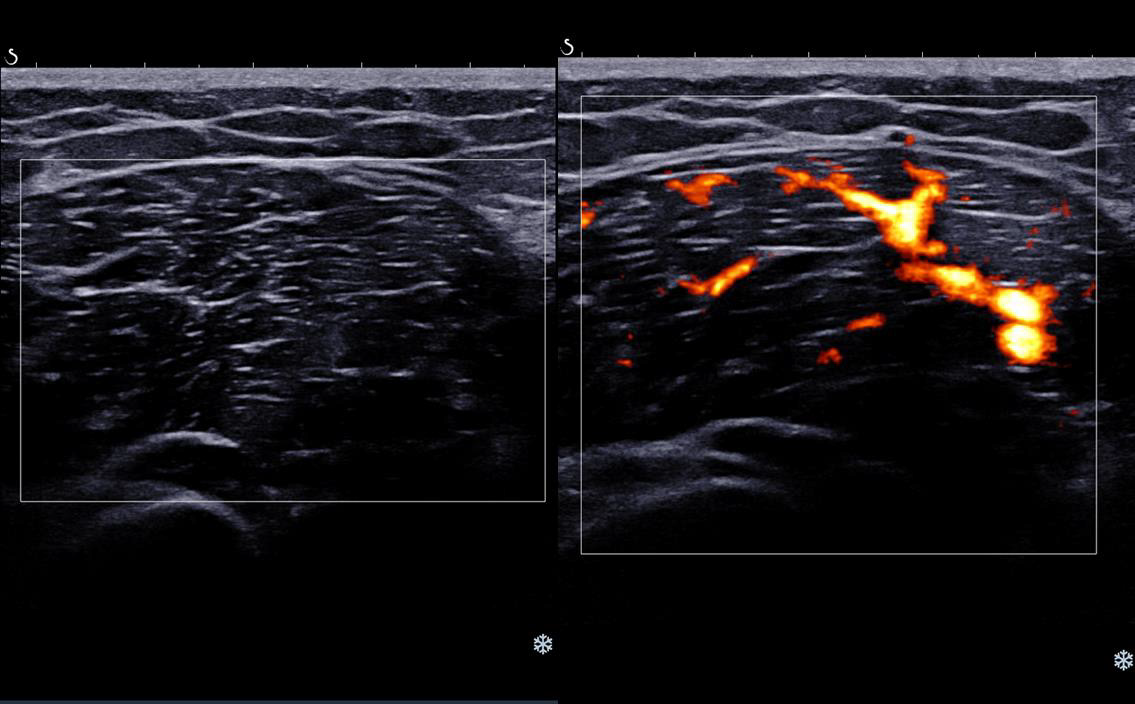
The power Doppler ultrasonic signal regions were different before and after exercise. (Figure 1) shows images of the same subject before (left) and after (right) exercise. The signal region significantly increased, indicating that muscle hyperemia significantly increased. Measurement of the signal regions from power Doppler ultrasonography before and after exercise was performed using Adobe Photoshop CS3 software. The signal regions were delineated. The value of a delineated region was read using the histogram interface of the software and expressed in pixels. Due to the difference in image magnification Z and the signal intensity gain value G, the signal regions of the medial head of the quadriceps muscle on power Doppler ultrasonography before and after exercise required correction using the following formula. The corrected power ultrasonic intensity was determined and expressed in pixels.

biomedres-openaccess-journal-bjstr

Figure 1: Comparison of the signal intensity on power Doppler ultrasonography before and after exercise in the medial head of the quadriceps muscle.
biomedres-openaccess-journal-bjstr

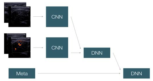
Figure 2: The convolutional neural network model (DNN: Deep Neural Networks. CNN: Convolutional neural network).

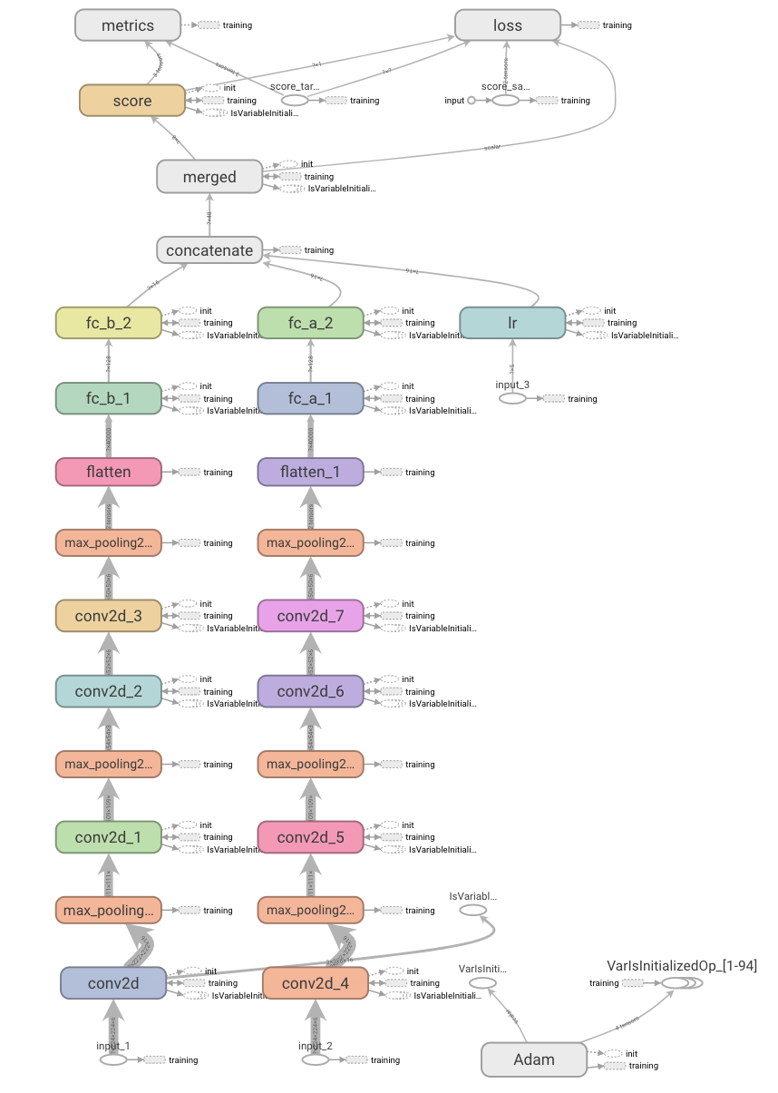
Multivariate linear regression: Multivariate linear regression analyses of the above measurement data were performed using SPSS 19.0 statistical software. Convolutional neural network model: Four known muscle strength images (2 before exercise and 2 after exercise) and participant information (gender, age, height, and body weight) were the inputs, and the knee extension peak torque was the output. A total of 160 groups of data consists of 132 healthy lower limbs and 28 quadriceps atrophy lower limbs from 80 volunteers was used as the training data set in this study, and the convolutional neural network was used for learning (Figure 2). The specific network structure included the use of two channels to extract features from the input data. The first channel extracted image features. The two groups of ultrasonic images before and after exercise were separately connected to one set of the convolution layer, which included 3 sections. The first section contained 1 3x3 16-kernel convolutional layer and 1 2x2 pooling layer. The second section contained 1 3x3 32-kernel convolutional layer and 1 2x2 pooling layer. The third section contained 2 3x3 64-kernel convolutional layers and 1 2x2 pooling layer. Feature extraction in ultrasonic images was performed using this set of convolutional layers to develop image features. The second channel extracted the 0/1 numerical features developed after binary processing of the personal information of the subjects. The numerical features and image features were connected to a neural network hidden layer containing 128 nodes for feature combination. The correlation between feature combination and fitting features was analyzed to obtain a final score, which served as the predicted knee extension peak torque. Algorithm design: The convolutional kernels of the two image convolutional channels were 3*3*16, 3*3*32, and 3*3*64, followed by a 128-neuron hidden layer. In addition, image features and numerical features were merged for feature combination through a 16-neuron hidden layer to obtain the final predicted score (Figure 3).
biomedres-openaccess-journal-bjstr

Figure 3: Design of the convolutional neural network algorithm.

Results

Forty-five men and 35 women were included among the volunteers. The basic information of the subjects is show. The quantitative ultrasonic technology parameters (muscle thickness, average muscle echo intensity, and corrected power ultrasonic intensity) and muscle strength parameter (knee extension peak torque) were introduced into SPSS 6.0 for data processing via multivariate linear correlation analysis. The dependent variable y, knee extension torque (Nm), was a continuous variable and conformed to a normal distribution. The independent variables were x1, muscle thickness (cm); x2, muscle average echo intensity (gray scale); and x3, corrected power ultrasonic intensity (pixel). The model R value was 0.708>0.4, and the adjusted R^2 was 0.492>0.1, indicating that the fitting degree of this model was relatively ideal, the goodness of fit was comparatively high. In the ANOVA table, the F value of the regression equation was 52.248. The probability of the significance test was 0 and was smaller than the significance level of 0.05. The coefficients were not 0 at the same time and the linear relationship between the explained variables and the overall explanatory variables was significant; therefore, a linear equation could be established. The coefficient table (Table 2) shows that the significance of all independent variables was smaller than 0.05, indicating that these 3 variables all had a significant linear correlation with the dependent variable. Furthermore, x1 and x3 showed positive correlations and x2 showed a negative correlation with the dependent variable, results that were consistent with the hypothesis. VIF values were all smaller than 2, indicating that the collinearity was not strong.

The final equation was obtained from this coefficient table.

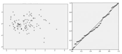
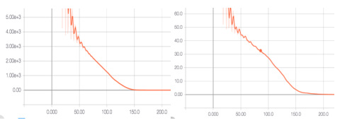
A residual analysis was performed. The P-P plot (Figure 4) showed that the source data and the normal distribution did not have significant differences and the residual met the requirement of a linear model. The scatter plot showed that these 3 points were evenly distributed on upper and lower sides of the vertical coordinate “0”, indicating that the data conformed to the model well. The products of pairwise multiplication among the three independent variables were introduced as interaction terms for multivariate linear regression. The coefficient table was then obtained. Except for x1, the significance was greater than 0.05, indicating no interaction among the independent variables. During the learning process of optimization iteration, MSE was selected as the loss function, and Adam was used as the optimizer algorithm to obtain the optimal value. After 200 rounds of iteration of 160 sets of data, the model results were obtained. After 200 rounds of iteration, the muscle strength accuracy rate was 93% and the range of the muscle strength accuracy was ±0.1 (Nm). By solving the model through the loss function, we hoped to achieve predicted and target values that were as close as possible using this model. After 200 rounds of iteration, the overall difference between the predicted and target values in the validation set was smaller than 0.01, indicating that the personal information and imaging information of the volunteers could be used to fit and approximate the real muscle strength data. The mean squared error (MSE) and mean absolute error (MAE) functions are shown in (Figure 5).
biomedres-openaccess-journal-bjstr

Figure 4: The P-P plot and the scatter plot.
biomedres-openaccess-journal-bjstr

Figure 5: The mean squared error (MSE) and mean absolute error (MAE) functions.

Discussion

The multivariate linear regression results not only confirmed that muscle thickness and average echo intensity correlated with muscle strength but also innovatively validated that the power Doppler ultrasonic parameters also had strong correlations with muscle strength, further supporting the clinical application of quantitative ultrasonic technology as a muscle strength testing method. The neural network algorithm of AI provides a more convenient alternative to manual extraction of ultrasonic data. Only a few seconds are required between the acquisition of basic information and ultrasonic images of subjects and obtaining the predicted torque; therefore, the method of manual labeling and calculations using the corresponding formula are omitted and the application range of ultrasonic imaging in muscle strength assessment is thus increased. The sample size in this study was small; however, a model with excellent accuracy was still obtained. Various errors in the manual extraction of ultrasonic image data highlight the advantages of AI. This model can be incorporated into the ultrasonic imaging instrument as a plug-in function. By inputting the basic information of a patient and examining the twodimensional ultrasound and power Doppler ultrasound images before and after exercise, the predicted knee extension peak torque can be obtained using the convolution neural network model. This method has the following advantages.

The anatomical positioning is accurate, the muscle strength of any muscle can be theoretically determined, patients do not experience trauma and pain, ultrasound is very popular and inexpensive, no consumable supplies are used, and AI deep learning can further increase the accuracy of predicted results. This study not only recruited healthy subjects but analyzed some quadriceps atrophy patients, which makes the methods and conclusions of this study having a better practical value in clinical applications. Regardless of decreased muscle thickness caused by disuse or pathological atrophy, reduced muscle quality caused by muscle disease or aging, or abnormal muscle recruitment caused by upper or lower motor neuron damage, both the linear regression equation and the AI model have significant applicability. The focus of future research may be on a more scientific and effective exercise program and scenarios in which no gold standard of muscle strength assessment exists. We believe that combining quantitative ultrasonic technology and the AI imaging recognition algorithm in muscle strength testing in clinical practice is feasible.

Conclusion

The successful establishment of the regression equation and the AI model in this study provide certain bases for using quantitative ultrasonic technology as a new method for muscle strength testing in clinical practice.


For more Articles on: https://biomedres01.blogspot.com/

Friday, July 5, 2024

A Cross-Sectional Study on the Acceptance of Covid-19 Vaccine


A Cross-Sectional Study on the Acceptance of Covid-19 Vaccine

Background

Since the beginning of the 20th century, the world has witnessed several crises which are epidemic and pandemic in nature. In the year 1918-1920, the Spanish Flu is popularly known as Influenza occurred and affected about one-third of the world population. In the 21st century, the SARS outbreak occurred in 2003, the Middle East Respiratory Syndrome (MERS) outbreak occurred in 2015, and the latest recent novel (2019-2020) Coronavirus (COVID-19). All these are known to have negatively affected the world economy. The current pandemic COVID-19 is caused by Severe Acute Respiratory Syndrome Coronavirus 2 (SARS-CoV-2) (WHO, Cucinotta, et al. [1,2]). It is an acute respiratory infectious disease which was originated in Wuhan, China in December 2019, and swiftly become a global threat affecting 220 countries (WHO, Helmy [1,3]). As of September 2021, there are more than 219 million cases (infected persons) and more than 4.55 million deaths recorded worldwide (WHO) [1]. In the African continent, there were about 4% cases of mortality, and Nigeria having 193,000 cases and 2,480 deaths (Worldometer [4]).

The pandemic has resulted in a devastating impact globally, which prompted the limit to a movement among other restriction policies to contain the pandemic (Ilesanmi [5]), as most countries strategy was to lessen the transmission of the disease, especially by non-pharmaceutical interventions (NPIs), such as enforcing hands sanitization, face masks policy, travel restrictions, social distancing, and complete or partial lockdowns (Ilesanmi [6]). So far, these interventions have not been able to curtail the spread of the disease, but they are effective strategies to minimize the spread if properly adhered to. Medical technologies have been put in place to prevent and cure the disease; among are affordable, safe, and effective antiviral vaccines and drugs. Despite the high mortality rate in the world as a result of COVID-19, there were no approved antiviral drugs and vaccines to specifically fight against SARS-CoV-2 (Ilesanmi [7]) till the end of November 2020. As of December 2020, the US Food and Drug Administration (FDA) granted an Emergency Use Authorization for critically ill COVID-19 patients (FDA; Beigel, et al. [8,9]).

Nonetheless, the WHO recommended that this is not effective for COVID-19 but can only suppress the intensity of the disease (Rochwerg, et al. [10]). Vaccines are interventions effectively used to reduce disease’s high burden globally. They are usually reliable and cost-effective public health interventions for saving millions of lives (Rodrigues, et al. [11,12]) from polio, yellow fever, measles, etc. Following the trend of the SARS-CoV-2 in the second, third, and fourth quarters of 2020 (Wu, et al. [13]) and the global pandemic declaration by the WHO in March 2020 (Cucinotta, et al. [2]) public and private stakeholders including scientists and pharmaceutical organizations have resulted to developing vaccines (Coustasse, et al. [14]). It is pertinent to note that as of January 2021, at least 85 vaccines have been subjected to preclinical trial in animals, and 63 vaccines passed the test and were subjected to clinical development in humans.

From these 63 vaccines, 43 were approved for phase I; from these 43, 22 were approved for phase II; from these 22, 18 were approved for phase III; from these 18, 6 were finally approved for early use though later restricted; from these 6,2 vaccines were approved for total use though one vaccine has been neglected (Coustasse, et al. [14]). Pfizer-BioNTech’s (BNT162b2) and Moderna’s (mRNA-1273) mRNA vaccines were approved for use, but Pfizer-BioNTech’s (BNT162b2) was widely accepted. With the news about the approval of COVID-19 vaccines, there is a tendency that the high surge of disease transmission will be minimized (Omer, et al. [15]). Nonetheless, there are hindrances to achieving the general acceptability of the vaccines, among the hindrances are the issues surrounding individual perception regarding the vaccine which is influenced by the level of socio-economic factors of an individual (such as education, age, culture), source of information, personal encounter, among all (Omer, et al. [15,16]), and more rampant in Africa and Nigeria (Ilesanmi OS [7]).

Vaccine hesitancy was recognized by the WHO Strategic Advisory Group of Experts (SAGE) as a “delay in acceptance or refusal of vaccination despite the availability of vaccination services” (Huo, et al. [17]). With the introduction of new health interventions, there are uprising issues regarding the interventions. For instance, the polio vaccination program in northern Nigeria was not accepted because of the wrong teachings of Islamic clerics (Jegede, 2007). This experience was also recorded in Ghana where community members did not comply with the de-worming interventions (Dodoo, et al. [18]). The major factor that was responsible for these rejections was a result of the lack of clarification (misunderstanding) on the interventions (Febir, et al. [19]).

It is therefore obvious that peoples’ knowledge of any infectious disease influences their acceptability of the interventions (vaccines) provided for tackling such disease. The acceptability of vaccine intervention is determined by three major factors: convenience [relative ease of access to the vaccine; physical availability of the vaccine; affordability and accessibility to the vaccine (Ilesanmi, et al. [20]), confidence [faith in the safety and efficacy of the vaccine; faith in the dynamics of healthcare delivery system; and faith in the policymakers (Zimmer, et al. [21]), and complacency [this is connected with diseases that are low risk and may not necessarily require vaccine; hence there are more negative acceptance towards the intervention of such diseases (Olaimat [22]).

Studies were conducted on the acceptance of citizens to the usage of COVID-19 vaccine, among the studies are Olaimat, et al. [22] on knowledge and information sources about COVID-19 among university students in Jordan; Pogue, et al. [23]on the influence of attitudes regarding potential COVID-19 vaccination in the United States; Malik, et al. [24] on the determinants of COVID-19 vaccine acceptance in the United States; Lazarus, et al. [25] on a global survey of potential acceptance of a COVID-19 vaccine in the United States; Coustasse, et al. [14] on the challenge of COVID-19 and vaccine hesitancy in the United States must overcome; El-Elimat, et al. [26] on the cross-sectional study of acceptance and attitudes toward COVID-19 vaccines in France and Jordan. Furthermore, Huo, et al. [17] conducted a study on the knowledge and attitudes about the Ebola vaccine among the general population in Sierra Leone; Febir, et al. [19] on the community perceptions of a malaria vaccine in the Kintampo districts of Ghana; Solís, et al. [27] on the COVID-19 vaccine acceptance and hesitancy in low and middleincome countries in Asia, Africa, and South America, Russia; and Olapegba, et al. [28] on COVID-19 knowledge and perceptions in Nigeria; Ilesanmi, et al. [5]on the perception and practices during the COVID-19 pandemic in an urban community in Nigeria.

The majority of these studies were conducted with the use of online respondents; they employed a cross-sectional approach of methodology; multinomial and binary logistic regression was found to be a dominant test of data analysis. However, the studies were most prevalent in the developed countries while scarcity of such studies in developing countries and Nigeria in particular. Because the level of acceptance of COVID-19 vaccines and the perception of COVID-19 differs among citizens of different countries, this study examines the acceptance of Nigerians to the usage of the COVID-19 vaccine.

Methods

This study employed a descriptive cross-sectional study in Nigeria. No consent was obtained as the data were collected and analyzed anonymously. A cross-sectional survey-based study was conducted in June 2021. Amid the global pandemic, data was further gathered from social media platforms. Also, online social media platforms (Facebook, WhatsApp) were used to recruit respondents (Olapegba, et al. [20,28]). With the high level of internet penetration in Nigeria which stands at 51.4% based on the nation’s population (Statista [29]), it is therefore justified that more citizens will be able to participate in the study. Respondents were encouraged to share the e-questionnaire with friends, contacts, or acquaintances. A sample of (n = 38) was employed to improve the clarity of the survey items. Data from the pilot sample was jettisoned as it was not used further for analysis. Categorical variables were presented as numbers and percentages, while continuous variables were presented as median [interquartile range].

The univariate analysis was performed using an independent Mann–Whitney U test for continuous variables and Chi-square test for categorical variables as appropriate. For analysis, responses to the attitudes section were combined. For example, both responses “strongly agree” and “agree” were combined in one category and both responses “strongly disagree” and “disagree” in one category. Before analysis, the independence of variables was analyzed using a correlation matrix. No multicollinearity was detected among predictor variables. To identify the factors that affect the acceptance of COVID-19 vaccines by Nigerians, binary and multinomial logistic regression were employed. The significance level [p <.05] was employed for affirming statistical decisions. Concerning the binary logistic regression model, the respondents were dichotomized as acceptable or not acceptable. The odds ratio (OR) values and the confidence intervals (95% CI) were calculated. The analysis was conducted using the Statistical Package for Social Sciences (SPSS Inc., Chicago, IL) version 23.

Results

Demographics

biomedres-openaccess-journal-bjstr

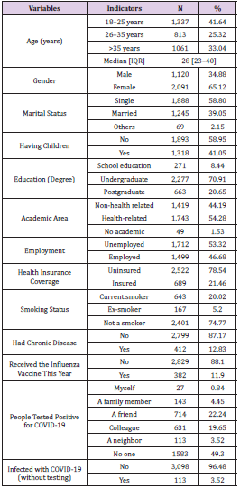
Table 1: Demographic details of study respondents (n = 3,211).

Note: Source: Authors’ work (2021)

biomedres-openaccess-journal-bjstr

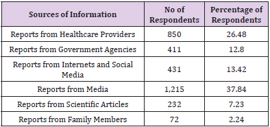
Table 2: Sources of information regarding COVID-19 vaccines.

Note: Source: Authors’ work (2021)

biomedres-openaccess-journal-bjstr

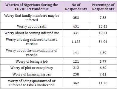
Table 3: Worries of Nigerians during the COVID-19 Pandemic.

Note: Source: Authors’ work (2021)

The study received 3291 submissions of which 3211 were complete and included in the final analysis. The median age of respondents was 28 years old and more than half of them were females (65.12%). More than half of the respondents were single (58.80%). About 70.91% had an undergraduate degree and more than half (54.28%) with health-related educational backgrounds. Besides, 46.68% of the respondents were employed and only 12.83% had chronic diseases (See Table 1). Less than 12% of the respondents received the influenza vaccine this year. About 0.84% of the respondents reported that they had tested positive for COVID-19. However, a minimal number of respondents (3.52%) stated that they might have been infected with COVID-19, but they did not verify it by laboratory test. As revealed in Table 2, about half (37.84%) of the respondents believed in the media reports while another significant percentage (26.48%) believed in the healthcare providers as a source of information about COVID-19 vaccines. About 13.42%, 12.8%, and 7.23% of the respondents believed the reports sourced from the internet and social media, reports from agencies, and reports from scientific articles respectively. During the COVID-19 pandemic, the respondents were worried about different issues (See Table 3). The major issue was the fear of being enforced to take a vaccine (34.94%), which is higher than the fear of death (13.42%) and fear of being quarantined or enforced to take a medication (11.28%). The fact that there are a sizeable number of respondents that have fear of taking vaccines, therefore, the acceptance of vaccines can be a bit negative. Hence, there is a need for serious sensitization among the general public.

Acceptance for COVID-19 Vaccines

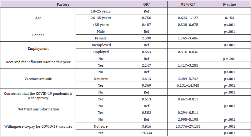
As earlier stated that there are a considerable number of respondents that have fear of taking vaccines, which may pose threat to the acceptability of the vaccines; Table 4 depicts the multivariate analysis (binary logistic regression) of independent factors that predicted the level of acceptance. The study found that older age categories (>35 years old) may not accept COVID-19 vaccines compared to younger age categories (OR = 0.487, 95 CI% = 0.328– 0.675, p< .001). Also, employed respondents (OR = 0.653, 95CI% = 0.516– 0.836, p <.001) may not accept COVID-19 vaccines compared to unemployed respondents. Respondents that believed in the conspiracy dynamics of COVID-19 pandemic (OR = 0.613, 95 CI% = 0.467–0.811, p< .001) and those that did not believe in the information (OR = 0.382, 95 CI% = 0.294–0.511, p< .001) may not accept the vaccine. On the other hand, males were more likely to have acceptance for COVID-19 vaccines (OR = 2.589, 95 CI% = 1.745–3.486, p< .001) compared to females.

biomedres-openaccess-journal-bjstr

Table 4: Predictors of acceptance for COVID-19 vaccines.

In addition, respondents who took the influenza vaccine this year were more likely to accept COVID-19 vaccines compared to those who did not take the influenza vaccine (OR = 2.147, 95CI% = 1.417–3.285, p = .001). Furthermore, respondents who stated that vaccines are safe in general were more likely to accept taking COVID-19 vaccines compared to those who stated that vaccines are not safe (OR = 9.369, 95CI% = 6.131–14.384, p <.001). Moreover, respondents who expressed their willingness to pay for COVID-19 vaccines have a high tendency of accepting the COVID-19 vaccines than those that did not show their willingness to pay (OR = 19.334, 95CI% =13.776–27.153, p <.001). The acceptance group will be dominated by people who did not believe in the conspiracy notion of COVID-19. Those who believe that vaccines are unsafe have the same ideology as those who are not willing to pay for the vaccine whenever it is available.

Discussion

This study examined the acceptance of Nigerians to the usage of COVID-19 vaccines and the factors influencing the level of acceptance. It was needed to unravel the doubtfulness regarding COVID-19 and its’ vaccines among the general public. The doubtfulness regarding the COVID-19 vaccine could reduce the efficacy of COVID-19 vaccines as soon as they are widely available nationwide (Olapegba, et al. [28]). Regarding the comparison of findings of this present study with the findings of previous studies on public acceptance and willingness to receive the COVID-19 vaccines nationwide, Nigeria is among the lowest countries with sufficient availability as the first batch of Oxford/AstraZeneca shots landed 2nd March 2021 and acceptance stands at only 1.23% as of 4th September 2021. While Africa as a whole only has 3% of its population vaccinated, Seychelles remains the highest vaccinated in Africa with 74.1% and Nigeria sitting at the 36th vaccinated even in Africa. From a study on the acceptance of COVID-19 vaccine across fifteen surveys covering Africa, Asia, Russia, South America, Russia, and the United States which targeted a total of 44,260 participants, it was revealed that there is a significantly higher level of acceptance towards taking COVID-19 vaccines with mean of 80.3%; median of 78%; range 30.1% in African countries, while mean of 64.6% and 30.4% in the United States and Russia respectively (Solís [27]).

Moreover, most western countries report relatively higher public acceptance. The acceptability level of vaccinations in Nigeria was far lower than global averages (Ilesanmi, et al. [20]). This was based on 440 respondents, and it was revealed that many individuals (67.30%) were aware of the prospective COVID-19 vaccine. This corroborates with the studies of Sani et al. (2016); Wang et al. (2018) which found a positive relationship between education and health awareness. In this study, factors influencing the acceptance of COVID-19 vaccines were analyzed with logistic regression. It was revealed that younger respondents were more likely to accept COVID-19 vaccines; this does not agree with the findings of El-Elimat, et al. [22,26] which found higher acceptance among older age categories. This may be as a result of different age distribution, and the fact that a country may be dominated by some categories of people (based on age), literacy level, and other factors that may influence the awareness of prospective health interventions. It is pertinent to note that alternate mediums of information dissemination could be employed for health interventions.

Examples are conventional media such as Radio, Television, and non-traditional media such as Facebook (Abdelhafiz, et al. [30]). COVID-19 pandemic as with other previous pandemics is associated with feelings of fear, anxiety, and worries (Olaimat [22]). Nonetheless, people are not fearful of getting infected or transmitting the disease to others, but they experienced economic and societal concerns because of the measures that were embarked upon by the governments to minimize the pandemic and halt human-to-human transmission of the disease (Dodoo, et al. [18]). These measures entail the enforcement of social distancing, lockdowns, curfews, self-isolation, borders’ shutdowns, school and universities closures, quarantine, and travel restrictions (Ilesanmi [5]).

In this current study, fear of taking vaccines and fear of death were most prevalent within the Nigerian population. This agrees with the findings of Holingue, et al. [31] among the US adults population which found that anxiety and fears of getting infected and died as a result of COVID-19 were linked to the increasing mental distress. In the study of French, et al. [32], the acceptance of the COVID-19 vaccine among college students in South Carolina was identified to be influenced by the information obtained from scientists (83%), followed by healthcare providers (74%), and then health agencies (70%). Nevertheless, contrary to this present study, information was not popularly disseminated by pharmaceutical companies in Nigeria. In the study of El-Elimat, et al. [25] in France, vaccination hesitancy and acceptance toward HBV and MMR vaccines were better when parents were informed through their healthcare providers. This seems more efficient than information sourced from the internet or a third party. However, it should be clearly stated that no matter the source of information, such information must be properly screened before absorbed.

Conclusion

This study aimed to examine the acceptance of Nigerians to the usage of COVID-19 vaccines and the factors influencing the level of acceptance. It was necessitated because of the level of doubtfulness regarding COVID-19 and its’ vaccines among the general public. An online cross-sectional study was conducted, achieved by the use of an e- questionnaire which was administered to respondents (Nigerians) in a form of an online survey with some particular emphasis on the COVID-19 vaccine acceptance. A total of 3291 respondents completed the survey, but 3211 responses were valid for data analysis and reporting, and logistic regression analysis was employed for data analysis. The Nigerian public COVID-19 vaccines acceptance was fairly low in Nigeria. The educated male respondents are most likely to accept the usage of the vaccine. Similarly, respondents who believed that vaccines are generally safe and those who were willing to pay for vaccines, after becoming widespread, were more likely to accept the COVID-19 vaccines.

However, those above 35 years old and respondents who are employed were not likely to accept the vaccines except been mandated by the employers. Moreover, respondents that believed in the rumors surrounding the dynamics of COVID-19 as well as those that do not have assurance in any source of information regarding COVID-19 vaccines, may not accept the usage of the vaccine. It was concluded that the most reliable sources of information regarding the COVID-19 vaccines were reports from the media and reports from healthcare providers. Organized interventions are essential by the authorities of healthcare providers to minimize the levels of doubt regarding COVID-19 vaccines, and advance approaches general acceptance. Further studies should be carried out to assess the awareness campaigns organized by both public and private stakeholders.

Take home message: Studies, reports and life experiences have established that COVID-19 is real in the developed and developing countries; therefore, the transmission is inevitable. Since the impact of COVID-19 on human life is fast killing, there is a need to employ vaccine to reduce the impact on human health. The available vaccine have been tested and passed through various stages such that it is fit for use.


For more Articles on: https://biomedres01.blogspot.com/
Thursday, July 4, 2024

Unusual Manifestation of Ectopic Human Chorionic Gonadotropin-Producing Cancers


Unusual Manifestation of Ectopic Human Chorionic Gonadotropin-Producing Cancers

Introduction

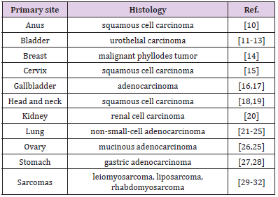
Elevated serum beta human chorionic gonadotropin (ß-hCG) in women generally indicates pregnancy, or less commonly, gestational trophoblastic disease, gonadal germ cell tumors, or extremely low levels from the anterior pituitary [1,2]. Elevation of hCG exist in several other states such as malignant tumors of nonendocrine origin (Figure1), and it has been suggested as possible tumor markers (Table 1). Some in vivo and in vitro studies have been shown that ß-hCG acts as an autocrine growth factor within tumor [3,4]. Elevated expression of ß-hCG may not be a reliable diagnostic marker, but it has been shown to serve as a strong indicator of poor prognosis for many non-endocrine tumors [1,3,4]. Elevated ß-hCG might manifest as gynecomastia in male patients or cause vaginal bleeding as initial symptoms in postmenopausal female patients [5-8]. These signs associated with high ß-hCG and subsequently increased level of estrogen may be attributed to paraneoplastic syndrome due to non-endocrine tumors. We purpose to understand the unusual sign of the ectopic ß-hCG producing malignancies as initial manifestation.

Gynecomastia in Males

Gynecomastia results from

1) Increased level of circulating estrogen, in particular estradiol,

2) Increased breast sensitivity to circulating estrogen, or

3) An altered balance of estrogen (stimulatory effect) and androgen (inhibitory effect).

Estrogen stimulates ductal epithelial hyperplasia, ductal branching and elongation, proliferation of the periductal stroma, and vascular distribution in the breast [5,6].

biomedres-openaccess-journal-bjstr

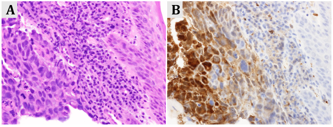
Figure 1: Stage IV gastric cancer in a postmenopausal women presenting with intermittent vaginal bleeding

(A) Microscopic image of gastric adenocarcinoma (hematoxylin/eosin staining, x400)

(B) Tumor cells were diffusely positive for human chorionic gonadotropin (hCG, x400).

Estrogen production in postmenopausal females and males results mainly from the peripheral conversion of androgens (testosterone, androstenedione and dehydroepiandrosterone sulfate,) by the action of the enzyme aromatase to estradiol and estrone [7,8]. The extra gonadal formation of estrogen occurs mainly in adipose tissue, skin and muscle. The patients with malignant tumors characterized by ectopic production of ß-hCG (Table 1) may be associated with excessive amounts of estradiol via the extragonadal metabolism of androgens.

biomedres-openaccess-journal-bjstr

Table 1: ÃŸ-hCG-producing tumors.

Vaginal Spotting in Females

The reproductive organs undergo progressive atrophy due to a reduced circulating estrogen and progesterone. This physiologic process of aging is also found at an endometrial level. Without the cyclic hormonal actions of the menstrual cycle, the endometrium during menopause becomes atrophic [9-18]. Non-physiologically increased estrogen level may cause postmenopausal bleeding. In a patient with unexplained vaginal spotting and no evidence of endometrial cancer or endometrial or cervical polyps, consideration should be given to the possibility of ectopic secretion of ß-hCG from other tumors.

Suspicion of Pregnancy in Premenopausal Females

In reproductive-aged women with elevated hCG, when pregnancy, either normal of abnormal (e.g. missed abortion, ectopic pregnancy)) is excluded, the malignancy should [18-24] be ruled out with imaging and surgical specimen immunohistochemically.

Comments

Pituitary hCG is naturally produced at extremely low levels in woman of reproductive age population. In peri- or postmenopausal state, levels of pituitary hCG increase because of the absence of feedback control by circulating estrogen and progesterone [2,25- 32]. Elevated ß-hCG level due to various cancers produce excessive amounts of estradiol by peripheral aromatase activation, which can manifest as gynecomastia in males or vaginal spotting in females. In either case estrogen and ß-hCG might be useful tumor markers during therapeutic management for checking recurrence of the disease, alongside other examination.


For more Articles on: https://biomedres01.blogspot.com/

Antimalarial Aloe Compounds

  Antimalarial Aloe Compounds Introduction Among the most prevalent diseases caused by protozoan parasites, malaria is caused by parasites o...