Friday, January 29, 2021

Potentials for Reducing Cancer Incidence in Nigeria

Potentials for Reducing Cancer Incidence in Nigeria


Opinion
Cancer genomics is changing oncology care and cancer detection rates in developed countries where genetic information can be used to improve care and management as well as inform decisions for personalized care for individuals owing to disparities in cancer genetics globally [1-3]. However, in some low and lower middle income countries, the battle against cancer is increasing [4]. For instance in Nigeria, cancer incidence and burden is growing with more than 115,000 new cases and over 70,000 deaths reported in 2018, with breast cancer as the leading type and cause of cancer mortality in the country [4,5]. Delays in detection, late presentation, poor access to treatment, poor prevention practice and knowledge and awareness all play significant roles in this increasing burdens while modifiable and unmodifiable risk factors for cancers have been identified [6-11]. Genetic predisposition account for some types of cancers originating from germ line mutations which increases the risk of developing especially ovarian, breast, colorectal, prostate and other cancers [12,13].

Although these hereditary cancers are inherited from parents, it does not always develop in their children. Because cancer cells undergo genetic changes which are detectable by high-throughput DNA sequencing, panel-based pathogenic gene screening through genetic testing, present many advantages for cancer management [14,15], but it is not without its challenges. This technology is however yet to be explored for reducing cancer burdens or for cancer care management in Nigeria. Genetic contributions to cancer have been studied widely in many populations and there are peculiarities in different populations; in the African descent. For instance, hereditary breast and ovarian cancers have been linked mostly to BRCA1 and BRCA 2 mutations in African and non-African populations [13,16], however, Africans are known to develop aggressive cancers and are at higher risks of deaths than other races. These variants of BRCA gene mutations in Nigerian population have been studied [19]. These unique genetic patterns and disparities should form the impetus for tailoring prevention, care and management for target populations with risk factors - like gender and ageing; which are not modifiable, and in turn greatly reducing the rising burden of cancers in Nigeria.

Breast and prostate cancers are two key cancer types that are prominent in Nigerians and the risk for both can be tested through genetic testing and potentially can overcome challenges of late presentation of patients which affects treatment outcomes and improve ways of managing and reducing cancer disease in Nigerian population. Mass screening for detection of new cases has helped previously in Nigeria and together with genetic testing and known modifiable risk factors linked to carcinomas in Nigerians [10,11,17,18], can reduce the risk of developing the disease. The tests are becoming available for home use and can detect early individuals who are at higher risk of cancer or those with a family history [19,20]. With adjustment of lifestyle, this can greatly reduce their risk of developing cancers and improve care and management for those affected in Nigeria.

A Practical Method to Predict the Incidence of Anterior Demodex Blepharitis-https://biomedres01.blogspot.com/2021/01/a-practical-method-to-predict-incidence.html

More BJSTR Articles : https://biomedres01.blogspot.com

Thursday, January 28, 2021

A Practical Method to Predict the Incidence of Anterior Demodex Blepharitis

A Practical Method to Predict the Incidence of Anterior Demodex Blepharitis


According to Medilexicon’s medical dictionary, the prevalence rate of Allergic conjunctivitis is around 10%. The causes include pollen, animal fur, contents of various eye drops, cosmetics, and dust mite tissue. Most of the symptoms can be resolved by the use of medication, however the symptoms of some patients can’t be relieved with medicine. One of the reasons is that the reproduction of mites and the rate of allergen creation is not altered by medication and Blepharitis becomes a chronic decease. Demodex blepharitis generally results from D. folliculorum and D. brevis [1-3]. The former infests the roots of eyelashes, becoming the leading cause of anterior blepharitis. The latter infests the meibomian gland, causing posterior blepharitis. The symptoms of inflammation caused by Demodex can vary from none to very diverse and are often hard to cure with medical treatment. Damage caused by the D. folliculorum includes direct damage to the hair follicle and the introduction of the two bacterium (Streptococci and Staphylococci) which cause hyperplasia stratum corneum, resulting in the instability of the eyelash leading to an increase in the feeling of foreign body sensation (FBS).
The two bacteria may also have links to skin conditions like rosacea [4]. It can be assumed that waste from the demodex and allergic reactions will cause the amount of patient discharge to increase. The proteins and excreta created by the D. folliculorum can cause itchiness, the fourth type of Allergic immune response [2]. Inflammation of the general surface layer of the eye can result in instability in the number of tears and may cause subjective feelings of dryness. In conclusion, the clinical symptoms can be categorized into FBS, discharge, itching, and dryness. Potential criteria for diagnosis of Demodex blepharitis are the following: Clinical history, Slit-lamp examination, and microscopic confirmation. Even though the third method, microscopic confirmation, can directly prove the existence of the condition, we cannot guarantee its nonexistence on patients with negative results due to being unable to check every eyelash. The purpose of this study is to find a way to increase the rate of diagnosis utilizing the third method, and also to find connections between the four patient symptoms (FBS, discharge, itching, and dryness) and the rate of infestation.

Patient Population and Selection
The study was conducted on 127 adult patients, composed of 29 males and 98 females, ranging from 58 to 96 years old, exhibiting at least one of the four symptoms.
Methods of Data Collection
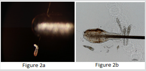
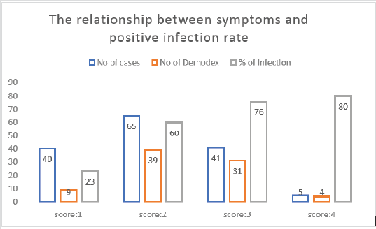
When patients voluntarily mentioned one of the four symptoms in clinic, the doctor included them as a candidate in this study. The doctor recorded the symptoms of each individual patient assigning a score equally depending on the number of symptoms (score1~4 as blue bar in Figure 1) and epilated three random eyelashes from every study participant’s upper eyelid on both sides. Sample preparation and identification of Demodex: The three epilated eyelashes from each participant’s eyelid, were placed individually by order on slides with one drop of Normal saline and covered with a cover slip. The samples were examined using a slit lamp and a microscope (25 X, Olympus SZX16 model) Figure 2a & 2b. All samples were evaluated within 10min of removal from the eyelid. If Demodex was discovered, it was identified by its order and the sample number was recorded (Figure 3). If all three samples were clear of demodex, then it was recorded as non-infested.

Figure 1: (The relationship between symptoms and positive infection rate: the blue bar represents the number of cases for each of the different score of symptoms. The red bar represents the number of positive demodex species found under microscopy in each group. The green bar represents the percentage of the red bar / blue bar (No. of cases with a positive finding in each group) / (No. of total cases in each group).
biomedres-openaccess-journal-bjstr
Figure 2: Observation of the D. folliculorum’s gathering around the hair follicle of an eyelash under slit lamp and microscopy (200~300Um).
biomedres-openaccess-journal-bjstr
Figure 3: The percent of Demodex found in the first and subsequent eyelashes is 52% (Blue area), 39%(Red area) , and 9%( Green area) in that order.
biomedres-openaccess-journal-bjstr
The positive diagnosed rate is 67%, with the average age of 72 years old (85/127). The clinical findings under slit lamp and microscopic findings are shown in figs.1a and 1b. The relationship between symptoms and positive infection rate is shown in Figure 1. The percent of Demodex found in the first and subsequent eyelashes is showed in Figure 3.

Clinically, Demodex infestation can cause many ocular diseases, i.e. the destruction of eyelashes, Meibimitis leading to trichiasis or enlarged glands, a hypersensitive reaction inducing lid margin inflammation, an unstable tear film or even superficial corneal vascularization, marginal infiltration, a phlyctenule-like lesion, superficial opacity, and nodular scarring [5,6]. At present, detection and counting of Demodex eggs, larvae and adult mites in epilated lashes under microscopy can make the final diagnosis under Slit-lamp examination (dandruff at the root of eyelashes [7,8] and clinical history (chronic blepharitis which is refractory to conventional treatments). However, a negative finding under microscopy cannot exclude the possibility of Demodex infestation. One limitation is that we can only see the D. folliculorum on top of the eyelash, and not those inside the sebaceous glands.

There is a strong correlation between a patient’s serum reactivity to Bacillus antigen (a bacterium from Demodex 9,10,11) and the presence of ocular rosacea. This is indirect evidence for Demodex infestation [9,10]. Due to the inability of the anterior blepharitis diagnostics to isolate substances produced by the demodex, we constructed a method that provides doctors with a highly plausible clinical testing process. The results of the experiment show that there is a high positive correlation between the symptoms and infestation rate. It is also found that around 90% of the infestations can be identified through the first two eyelashes inspected, but there is still a chance of it not being accurate. If the patient’s symptoms and signs are highly suspect for the diagnosis of demodex blepharitis, it is advisable to repeat the process to increase the diagnostic accuracy.

Demodex mite plays an important role in the occurrence of a series of ocular surface diseases such as Demodex blepharitis, meibomian gland dysfunction, conjunctival inflammation, and corneal lesions. Ocular infestation has a close relationship with the systemic infestation. Further studies are needed for developing easy and sensitive diagnostic methods and more effective and specific treating regimens.

Clinical and Bacteriological Diagnosis of Foot-rot in Beef bulls in Basra-https://biomedres01.blogspot.com/2021/01/clinical-and-bacteriological-diagnosis.html

More BJSTR Articles : https://biomedres01.blogspot.com

Clinical and Bacteriological Diagnosis of Foot-rot in Beef bulls in Basra

Clinical and Bacteriological Diagnosis of Foot-rot in Beef bulls in Basra


Introduction
Foot-rot is an acute and extremely infectious bacterial disease that generates lesions on the subcutaneous tissues and skin of the interdigital space of ruminants [1]. The disease is caused by Fusobacterium necrophorum. A rod shaped gram-negative aerotolerant anaerobe opportunistic bacterium; that presents normally in the feces and rumen of cattle and in their environment [2]. Fusobacterium necrophorum has the ability to cause considerable hepatic abscessation in cattle, sheep and camel [3]. Two subspecies of this bacterium were recognized; F. necrophorum sub-species necrophorum F. necrophorum and F. necrophorum sub-species fundulforme. The sub-species necrophorum is detected more frequently in foot-rot infections than the sub-species funduliforme as a result of its intensive virulence [4]. Several secreted products and/or toxins have involved as virulence factors. Haemagglutinin, endotoxin and leukotoxin [5], haemolysin and adhesion [2] are the main factors assisting the ruminal localization and liver invasion. Among these toxins, “endotoxin” and “leukotoxin” are thought to be more effective than others in overcoming the hosts defense mechanism to induce the infection [2,5]. Many attempts for providing a protective immunity against F. necrophorum by using toxoid, whole cell bacterin or other cytoplasmic compositions have been investigated. Unfortunately, none of the prototype immunogens has been introduced a satisfy immunity against fusobacterial infection. Therefore, the control of foot-rot has mainly based on the using of the anti-microbial therapy [6]. When there is injury to the skin of the interdigital space especially where the cattle are located on muddy and contaminated areas, F. necrophorum can invade causing the disease. Then the infected cattle will further spread the organism to the environment and to other susceptible [7]. The high cost of treatment and sharp decrease of weight gain leading to heavy economic losses in cattle industry [8]. Cattle with severe hooves infection might require to be euthanized, while the lame bulls could not breed accordingly. The economic impact of this disease will be minimized through appropriate preventive methods and early treatment. Minerals deficiency (Zink, Selenium and Copper) could probably increase the incidence of foot-rot [9].
Little is known on the pathogenicity of F. necrophorum in foot-rot infection with no single study has been clarified the sub-species of this organism that presents in hooves in Basra. Therefore, in the current study, we used clinical, bacteriological and biochemical analysis on hoof samples from foot-rot infected bulls to identify the F. necrophorum, and to study its role on the blood parameters.

Methodology
The current study was done to investigate the causes of hooves lesions of bulls in a private beef cattle farm that located at the south of Basra province during October 2017. Thirty-one beef bulls aged between 2 - 2.5 years were found to have hoof lesions, all bulls were inspected clinically, and all the affected animals were treated accordingly. Blood and hoof swab samples were collected and analyzed as following:

Hematological Analysis
Jugular blood was collected in EDTA tubes for Complete Blood Count (CBC) analysis. The samples were analyzed manually in clinical pathology laboratory, department of internal and preventive medicine, college of veterinary medicine / university of Basra. The examined parameters were: Red Blood Cell Counts (RBCc), Hemoglobin (Hb), Packed Cell Volume (PCV), Mean Corpuscular Volume (MCV), Mean Corpuscular Hemoglobin Concentration (MCHC), total White Blood Cell Count (WBCc), Lymphocyte Count (Lymp), Segmented Neutrophils Count (S Neut), Monocyte Count (Mono), Eosinophil Count (Eosin) and Basophiles Count (Baso).

Bacterial Isolation and Identification
Hoof scrapings of the thirty-one lame bulls were collected anaerobically. Samples were cultured directly on Macconkey and Blood agar culture in CO2 jar at 37°C for 48 hours in Central Research Unite, College of Veterinary Medicine / University of Basra.
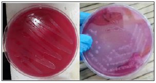
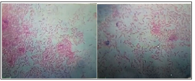
Initial bacterial determination based on the characteristic morphology of the growing colonies and the organism. When the colonies irradiated with UV light, a greenish fluorescence appearance can be noticed. Butyric acid odor also can be smelled. Microscopically, characteristic rod shape gram negative pleomorphic morphology of the bacterium was noticed. In addition, the identification was appended by the susceptibility to metronidazole and kanamycin on primary inoculated agar plate. To confirm the identification of F. necrophorum, a marked β-hemolysis on blood agar enriched with 5% sheep blood in conjugation with resistance to vancomycin and susceptibility to polymyxin and the specific biochemical tests were applied as described by [10].

Statistical Analysis
All data were analysed using JMP® 11. NC: SAS Institute Inc. software version. The data were considered significant at (p<0.05). Pair Student’s “t” test was applied to determine the differences.

Results
Clinical Examination
The symptoms of the disease were recorded as following:
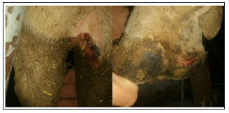
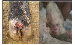
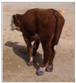
Sudden onset of lameness accompanied with decrease appetite (Table 1). The bulls were often only touching their toe to the ground due to sever painful lesions. More than one limb was often infected, commonly the fore-limbs. The tissues of the interdigital space were reddish and swollen that consequently leads to separate a part of the hoof. Skin between toes of some infected bulls develops a crack (Figures 1-3). Moderate to severe fever with significant increase of respiratory and heart rates (Table 1). There was a marked foul smell.

Table 1:Clinical and bacteriological examination results of the infected bulls.
biomedres-openaccess-journal-bjstr
Figure 1: Case of infected hoof showing: Crack, swelling and dead tissues in the interdigital space.
biomedres-openaccess-journal-bjstr
Figure 2: Case of infected hooves showing: swelling, necrotic area, crack and dead tissues in the interdigital space.
biomedres-openaccess-journal-bjstr
Figure 3: Case of infected bull showing: Signs of restlessness and lameness.
biomedres-openaccess-journal-bjstr
Isolation and Identification of F. necrophorum
Of thirty-one swabs taken from symptomatic foot-rot bulls, twenty-nine were found to be positive, according to the morphology of the colonies (Figures 4-6) and the biochemical characteristic of the organism (Table 2). The colonies appearance was: circle in shape, slightly elevated with smooth edge, moveable when touched has a white buff with a waxy appearance, occasionally the color changed to light yellow. Marked β-hemolysis zoon around the colonies of F. necrophorum is seen on the blood agar. Nevertheless, two swabs that have collected from the infected hooves gave negative results to the microbiological examination.
Figure 4,5: A characteristic morphology of F. necrophorum culture colonies on Macconkey agar.
biomedres-openaccess-journal-bjstr
Figure 6: Photomicrograph of a Gram stain bacterial smear showing a characteristic Gram-negative rode shape F. necrophorum.
biomedres-openaccess-journal-bjstr
Complete Blood Count
The mean of hemogram parameters that recorded in the current study were with the normal values, while marked changes in total white blood cell, monocytes and lymphocytes percentages were recorded (Table 3).
Table 2: Results of the biochemical characterization of F. necrophorum.
biomedres-openaccess-journal-bjstr
Table 3: Complete Blood Count Analysis Results of the Infected Bulls (Means ± S.E.).
biomedres-openaccess-journal-bjstr
Discussion
Foot-rot is a major problem to animal welfare that leading to reduction in productivity [8]. Fusobacterium necrophorum is the main etiology of foot-rot, and an opportunistic bacterium that could causes other conditions such as: hepatic abscesses [3], oral and laryngeal coccobacillus’s [2]. Understanding the pathogenicity of F. necrophorum and which strain are present in endemic footrot areas, are of value for satisfy treatment, control and eradication of the disease. Little information is available of the genetic variety of F. necrophorum in Iraq. To our knowledge, the current study is the first for identification of F. necrophorum on hooves swaps that have been collected from infected bulls in Basra province. The high absolute (94%) positive reporting of F. necrophorum from affected toes highlights it’s important role in foot-rot infection, and supporting the findings of previous researches that have been done by [2, 4, 5, 7, 8, 11-15]. Moreover, F. necrophorum is almost presents on the toes of the lame cattle.

In this study, we were focusing on F. necrophorum as a primary cause of bovine foot-rot. Detection only a single variation from any individual sample could be indicated that the pathogen variants are very specialized and there was a lack in train diversity as well as the little number of foot-rot specimens. Further molecular analysis can reveal more details of this pathogen. Fusobacterium necrophorum population in rumen is greater in grain feed than forage feed animals. Consequently, to rumenitis and ruminal acidosis of grain-fed cattle, the bacteria gain access to the portal circulation then localized in liver causing abscessation [5]. In this study, F. necrophorum was detected from swabs have taken from the hoof of bulls. This finding proposes that the organism might be transmitted to and from the mouth and rumen of cattle to the beef farm, so far by an un-characterized pathway. In our research, all the examined bulls were shown to have a sever hoof lesion which could be as a role of leukotoxin. In cattle, it is possible that the leukotoxin that secreted from bovine F. necrophorum plays a key role in the creation of necrotic infections [16-18].

A complete blood count used to be suitable as a screening assessment for underlying certain illness such as infectious diseases or anemia [19,20]. To our knowledge, this work is the first study to demonstrate the effect of foot-rot infection for local strain of F. necrophrum on beef cattle in Basra and relationship of this bacterium on hemogram and leukogram changes after footrot infection. In the current study, the hemogram parameters that recorded in all infected bulls were within the normal range suggesting that there were no severe effect of the virulence of F. necrophorum on the hemogram parameters during the acute phase of the disease. Unfortunately, we did not collect blood samples prior the onset of foot-rot infection and hemogram values could be higher before infection. However, chronic Fusobacterial infection such as liver abscessation could induce a significant alteration in blood parameters of beef bulls [21].

On the other hand, the increase in leukogram values especially monocytes and lymphocytes suggest an inducing of inflammatory reaction which needs further production of monocytes to play its role as phagocytes as a response to F. necrophorum infection or due to the bad management, environmental factors and previous infection that subsequent contribute to develop of foot-rot in the beef bulls [1].

Conclusion and Recommendation
This study reports the initial data on F. necrophorum in beef cattle flock with different clinical aspects of foot-rot in Basra province regarding virulence of the organism. Fusobacterium necrophorum is often found on the hooves of lame beef cattle in Basra. The risk of this organism in lame beef herds might be of value for management of other susceptible animals as F. necrophorum is believe it to be implicated in foot-rot likewise as reported in [13]. Therefore, further studies should be done for molecular analyze to the genetic variety of this organism. Study the rule of bacterial toxicity and the virulent factor of the bacterium to understand the pathogenesis of foot-rot also needed. Additionally, highlight the importance or foot-rot as a threat of animal heath welfare and its economic losses in beef cattle.

Molecular Conformation Correlation to Activity Against Herpes Simplex Virus of (E)-5-(2-Bromovinyl)-2’-Deoxycytidine and 5-Methoxymethyl-2’-Deoxycytidine Analogs: Short-Review-https://biomedres01.blogspot.com/2021/01/molecular-conformation-correlation-to.html

More BJSTR Articles : https://biomedres01.blogspot.com

Molecular Conformation Correlation to Activity Against Herpes Simplex Virus of (E)-5-(2-Bromovinyl)-2’-Deoxycytidine and 5-Methoxymethyl-2’-Deoxycytidine Analogs: Short-Review

Molecular Conformation Correlation to Activity Against Herpes Simplex Virus of (E)-5-(2-Bromovinyl)-2’-Deoxycytidine and 5-Methoxymethyl-2’-Deoxycytidine Analogs: Short-Review


Introduction
The antimetabolites (E)-5-(2-bromovinyl)-2’-deoxycytidine (BrVdCyd) (1) and 5-methoxymethyl-2’-deoxycytidine, MMdCyd (2) are selective and potent antiherpes agents with low cytotoxicity [1,2,3]. The antiviral activity of BrVdCyd (1) and MMdCyd (2) is influenced by the cytidine/deoxycytidine (Cyd/dCyd) deaminase and deoxycytidilate (dCMP) deaminase content of the cell lines used for antiviral assays [1]. When deamination is prevented, both (1) and (2) are potent inhibitors of herpes simplex virus type 1 (HSV-1). The IC99 of BrVdCyd (1) (concentration required to reduce the yield of infectious virus obtained 72 h after infection by 99% relative to control cultures) was 1.6 μM when (1) was used in combination with tetrahydro-deoxyuridine (H4dUrd; an inhibitor of both dCyd and dCMP deaminases) (1). BrVdCyd (1) is also a good inhibitor of varicella zoster virus (VZV) replication [1]. Previous studies have shown that resistance to deamination for cytidines can be accomplished by structural modifications of the cytidine molecule [3-8]. Therefore, systematic investigations on the development of 5-substituted deoxycytidine analogs of (1) and (2) resistant to deamination were initiated.

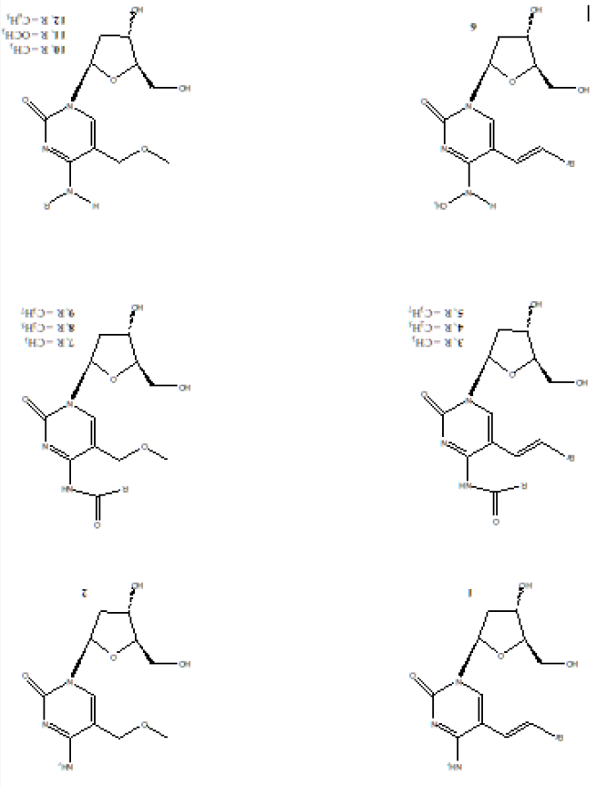
The rationale is that deoxycytidine analogs resistant to deamination would retain selectivity and metabolic stability thus simplifying HSV-1 treatment regimens. Therefore, N4-substituted derivatives of (E)-5-(2-bromovinyl)-2’-deoxycytidine (BrVdCyd) and 5-methoxymethyl-2’-deoxycytidine (MMdCyd) were synthesized to confer resistance to deamination, to improve delivery of the antimetabolite and to determine the bulk tolerance of the viral kinase to the N4-substituents of the cytosine moiety. The molecular conformation in solution was determined by NMR spectroscopy and the activity against herpes simplex virus type 1 (HSV-1) was determined using A549 cells. Compounds (3) and (4) were also found to be 100 times more potent and selective inhibitors of the varicella zoster virus (VZV) compared to the widely used antiviral drug acyclovir [9]. Chemical structures for compounds (1 - 12) are shown in Figure 1.

Figure 1: Chemical structures of compounds (1 – 12). BrVdCyd (1), MMdCyd (2), N4-acetyl-BrVdCyd (3), N4-propanoyl- BrVdCyd (4), N4-butanoyl-BrVdCyd (5), N4-methyl-BrVdCyd (6), N4-acetyl-MMdCyd (7), N4-propanoyl-MMdCyd (8), N4- butanoyl-MMdCyd (9), N4-methyl-MMdCyd (10), N4-methoxy-MMdCyd (11), N4-phenyl-MMdCyd (12).
biomedres-openaccess-journal-bjstr
Experimental
Antiviral Activity
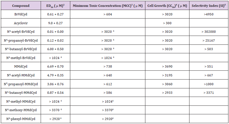
The effectiveness of compounds (3-12) as inhibitors of HSV- 1 (McIntyre strain) replication was determined by the plaque reduction assay using A549 cells. The parent compounds BrVdCyd (1) and MMdCyd (2) were used as a positive control. N4-Acetyl- BrVdCyd (3) was significantly more active (ED50 = 0.01 μM) than its parent compound BrVdCyd (1) (ED50 = 0.61 μM), N4- propanoyl-BrVdCyd (3) was a good inhibitor of HSV-1 replication (ED50 = 0.12 μM) and N4-butanoyl-BrVdCyd (4) (ED50 = 6.00 μM) was a poor inhibitor compared to their parent compound (1). N4- Methyl-BrVdCyd (5) was devoid of activity (ED50 > 1024 μM). All compounds have low cytotoxicity and can be considered essentially nontoxic as measured by the CC50 (cytotoxic concentration required to reduce cell growth by 50% using confluent monolayers of A549 cells). Table 1 shows the antiviral activity data for all compounds with acyclovir’s activity added for comparison.

Table 1: Antiviral activity of the compounds listed against HSV type-1 in A-549 cell line1.
biomedres-openaccess-journal-bjstr
Note:
a) HSV-1 McIntyre strain was used. Virus input was 50 PFU.
b) ED50: Inhibitory concentration required to reduce viral plaques by 50% (mean + SD, n = 12)
c) MCC: Minimum cytotoxic concentration that causes a microscopically detectable alteration of cell morphology.
d) CC50: Cytotoxic concentration required to reduce cell growth by 50%.
e) SI: Ratio of CC50 /ED50.
f) Highest concentration tested.
Conformational Analysis
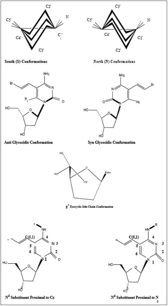
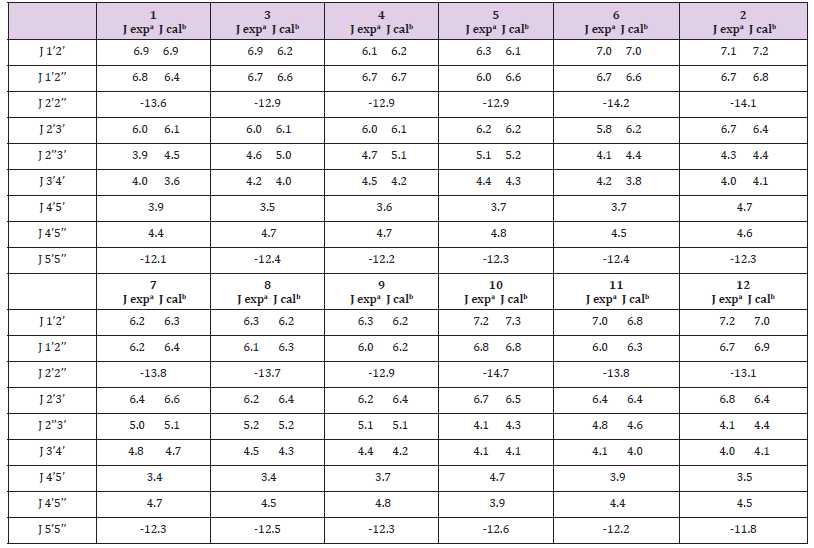
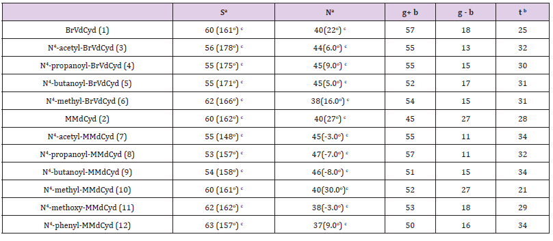
The in-solution conformation of compounds (1-12) was determined by NMR spectroscopy using D2O as a solvent. Four different conformational aspects were under consideration (Figure 2): The North/South deoxyribose ring pseudo-rotational conformation, the Syn/Anti glycosidic conformation, the exocyclic side-chain conformation (g+, g- and t) and finally orientation of the N4-substituent (proximal to C5 or proximal to N3 of the pyrimidine ring). The proton-proton coupling constants are given in Table 2; conformational parameters are summarized in Tables 3 & 4. The four conformational modes under study are shown in Figure 2 (structures for N4-substituted BrVdCyd and its analogs are provided). The conformation of the deoxyribose sugar ring was obtained form the relationship between the proton-proton coupling constants and the pseudo-rotational properties (Table 3) of the ring using the computer program PSEUROT [10]. The population of the three rotamers (g+, g- and t) about the exocyclic C(4’)-C(5’) bond (Table 3) was determined from the J4’5’ and J4’5’’ coupling constants [11,12].

Figure 2: The four conformational modes in pyrimidine deoxyribonucleoside analogs discussed in this study.
biomedres-openaccess-journal-bjstr
Table 2: Vicinal coupling constants (experimental and calculated in Hz) of BrVdCyd (1), N4-acetyl-BrVdCyd (3), N4-propanoyl- BrVdCyd (4), N4-butanoyl-BrVdCyd (5), N4-methyl-BrVdCyd (6), MMdCyd (2), N4-acetyl-MMdCyd (7), N4-propanoyl-MMdCyd (8), N4-butanoyl-MMdCyd (9), N4-methyl-MMdCyd (10), N4-methoxy-MMdCyd (11) and N4-phenyl-MMdCyd (12)
biomedres-openaccess-journal-bjstr
a: J experimental; precison 0.1 Hz.
b: J calculated using PSEUROT with tm =360 C
Table 3: Conformation populations (%): South (S) and North (N) furanose ring conformers and the three rotamers (g+, g- and t) of the exocyclic C(5’) side chain of BrVdCyd, MMdCyd and their N4 substituted analogs.
biomedres-openaccess-journal-bjstr
Note:
a) In PSEUROT calculations, μm was constrained to 36.0o. The rms deviation for each calculation was: BrVdCyd: 0.193; N4- acetyl-BrVdCyd: 0.250 E; N4-propanoyl-BrVdCyd: 0.227 E; N4- butanoyl-BrVdCyd: 0.179 E; N4-methyl-BrVdCyd: 0.175 E; N4- acetyl-MMdCyd: 0.204 E; N4-propanoyl-MMdCyd: 0.166 E;N4-butanoyl-MMdCyd: 0.181 E; N4-methyl-MMdCyd: 0.112 E; N4- methoxy-MMdCyd: 0.145 E; N4-phenyl-MMdCyd: 0.202 E.
b) C5’ Exocyclic orientation at 37oC.
c) Numbers in brackets are the calculated pseudo-rotational phase angles PS and PN
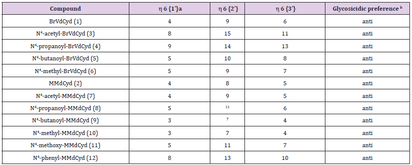
The syn/anti glycosidic preference (orientation of the pyrimidine ring relative to the deoxyribose moiety) defined by the torsion angle O-C(1’)-N(1)-C(2) was determined by nuclear overhauser enhancement (nOe), the enhancement of deoxyribose protons H1’, H2’ and H3’ were observed on pyrimidine H6 using the method of Davies [13] and the data are summarized in Table 4. The position of the N4-substituents (proximal to C (5) or proximal to N (3)) was elucidated by observing the nOe of the N4-substituent protons or lack of it on C (5,1) and C (5,2) olefinic (vinyl) protons in BrVdCyd analogs and in the case of MMdCyd analogs, the nOe of the N4-substituent protons or lack of it on C (5,1) methylene and C (5,3) methoxy protons [14].
Table 4:Syn/anti glycosidic preference of BrVdCyd, MMdCyd and their N4 substituted analogs.
biomedres-openaccess-journal-bjstr
Note:
a) The common notation used for nOe is ηi {s} which indicates that this is the nOe of nucleus i when nucleus {s} is saturated.
b) Glycosidic preference refers to a dynamic solution equilibrium that is biased towards either syn or anti. For syn orientations, H6 is closest to H1’ and the nOe to H6 will be mainly from H1’. For anti-orientations, the nOe to H6 will be mainly from H2’ and H3’

Results and Discussion
Deoxyribose Ring Conformation
The four major modes of conformational flexibility for pyrimidine 2’-deoxyribonucleosides are shown in Figure 2. The conformation of the deoxyribose moiety is important in determining the biological activity of 5-substituted 2’-deoxyribonucleosides against herpes simplex virus and varicella zoster virus [2,3,5,15]. All compounds (1 - 12) displayed South/North equilibrium for the deoxyribose conformation in the range from 53/47 to 63/37.
C (5’) Exocyclic Side Chain Conformation
Molecular conformation studies by X-ray crystallography (solid state) and NMR spectroscopy (in D2O) indicate that the conformation of the 5’-exocyclic side chain [γ torsion angle C (3’)- C (4’)-C (5’)-O (5’)] is important in determining the activation of 5-substituted pyrimidine-2’-deoxynucleosides by HSV- induced thymidine kinase (HSV-TK). The g+ conformer seems to be the preferred orientation required by HSV-TK; whereas the t conformer appears to be an unfavored conformation [3,7,9,15]. All compounds (1 - 12) have a predominant g+ rotamer for the C (5’) exocyclic side chain (Table 3).
Syn/Anti Glycosidic Conformation
The syn/anti glycosidic equilibrium in all compounds (1-12) is biased towards the anti-region (Table 4).
N4-Substituent Orientation
The in-solution orientation of the N4-acyl substituent in anti HSV-1 active compounds of both series BrVdCyd (1) and MMdCyd (2) (compounds 3, 4, 5, 7, 8 and 9) is proximal to pyrimidine C (5). Whereas the orientation of the N4 substituent in compounds 6, 10, 11 and 12 all of which showed no anti HSV-1 activity is proximal to pyrimidine N (3). The aforementioned results were established by nOe experiments on all ten derivatives of BrVdCyd (1) and MMdCyd (2) namely compounds 3 - 12.

Conclusion
In considering the four conformational modes of the compounds under study, we found that those analogs which had no activity against HSV-1 have three of the four modes in common with those analogs which were active against HSV-1, namely the deoxyribose ring conformation (predominantly south), the exocyclic sidechain (predominantly g+) and the glycosidic bond conformation (predominantly anti). The one difference between analogs which are active against HSV-1 and analogs which are not is the N4- substituent orientation. Anti-HSV-1 active analogs had the N4- substituent oriented proximal to pyrimidine C (5) whereas analogs lacking any activity against HSV-1 had the N4-substituent oriented proximal to pyrimidine N (3). Therefore, it is safe to conclude that an N4-substituent oriented proximal to pyrimidine N (3) prevents in some way an analog from exhibiting activity against HSV-1. It is obvious that the potency trends in the two series N4-acyl-BrVdCyd and N4-acyl-MMdCyd analogs run in opposite directions. While in the one series, the analog N4-acetyl-BrVdCyd is the most active among three N4-acyl analogs, it is the N4-butanoyl-MMdCyd that is most active among its three N4-acyl analogs.

In general, we conclude that the N4-acyl substituents conferred resistance to deamination in MMdCyd analogs with N4-butanoyl- MMdCyd being the slowest to undergo hydrolysis of the N4- butanoyl group and eventual deamination, hence shows the greatest antiviral activity. Steric bulk in N4-acyl MMdCyd substituents did not seem to affect ease or rate of phosphorylation by HSV-1 induced thymidine kinase. We also conclude that N4-acyl substituents conferred resistance to deamination in BrVdCyd analogs yet it appears that bulk tolerance reverses the anti HSV-1 trend seen earlier in MMdCyd analogs. N4-Acetyl-BrVdCyd shows resistance to deamination and an overall tolerable bulk. As the substituent grows bulkier to N4-propanoyl and N4-butanoyl, anti HSV-1 activity is less especially with N4-butanoyl-BrVdCyd perhaps due to docking and rate of phosphorylation by HSV-1 induced thymidine kinase.

Presumed Orbital Infarction Syndrome after Functional Endoscopic Sinus Surgery (FESS)-https://biomedres01.blogspot.com/2021/01/presumed-orbital-infarction-syndrome.html

More BJSTR Articles : https://biomedres01.blogspot.com

Antimalarial Aloe Compounds

  Antimalarial Aloe Compounds Introduction Among the most prevalent diseases caused by protozoan parasites, malaria is caused by parasites o...