Wednesday, October 25, 2023

Jaundice Clinical Manifestation and Pathophysiology: A Review Article


Jaundice Clinical Manifestation and Pathophysiology: A Review Article

Introduction

Jaundice is when clinically there is an increase in the amount of bilirubin in the serum rising above 85mmol/l (5mg/dl). When in utero, unconjugated bilirubin is cleared in the placenta to produce cord serum bilirubin of approximately 35mmol/L (2mg/ dl). After birth, jaundice is a reflection of the bilirubin present in the liver, the rate of hepatic excretion and the ability to bind to serum proteins to retain the bilirubin present in the plasma. Many variations in individual responses to bilirubin load prevent specific levels of psychological jaundice [1]. Therapy has proven that the benefits outweigh the harm. The physiological pattern of jaundice also varies with other factors such as prematurity and ethnicity. In hemolytic jaundice, serum bilirubin exceeds physiologic levels and can be caused by sepsis and inherited hemolytic diseases such as glucose 6-phosphate dehyrogenase (G6PD) deficiency, ABO and Rh isoimmunization [2]. Prolonged jaundice refers to jaundice that persists beyond the first two weeks of long neonatal life, and is caused by other diseases, including lateonset jaundice of the breast, congenital hypothyroidism and rare inherited disease conditions. Infants with breast milk jaundice have prolonged levels of unconjugated bilirubin in other healthy infants, and glucuronidase b in breast milk appears to be an important factor in this condition. There are two types of neonatal jaundice associated with breastfeeding, the first known as lactational jaundice associated with treatment of the breast which results in dehydration and causes the jaundice to become more intense [3]. The second is breast milk jaundice which is associated with prolonged jaundice in infants with the first two weeks of life. One or more substances such as enzymes (glucoronidase), 3 alpha, beta and 20 pregnanediol in breast milk although to be possible cause of breast milk jaundice. Jaundice is influenced by several factors, namely dehydration, weight loss after birth, bleeding, cephalohaematoma, contusions, babies in mothers with diabetes, acidosis, apixia, and gastrointestinal obstruction [4].

Discussion

Clinical Manifestation and Etiology

Patients with jaundice may not experience any symptoms, although some present with a life-threatening condition. Patients who present with acute illness, which is usually due to infection, may present with fever, chills, abdominal pain, and flu-like symptoms. In these patients, skin discoloration may not be their chief complaint. Patients with non-infectious jaundice may complain of weight loss or pruritus. Abdominal pain is a symptom that usually appears in pancreatic or biliary tract carcinomas. Occasionally patients present with jaundice and accompanying extrahepatic manifestations of liver disease

Jaundice can be caused by a number of things, including [4,5]:

a. Haemolysis due to ABO or Rh isoimmunisation, resulting in G6PD. deficiency

b. Hereditary spherocytosis, drugs

c. Sepsis - septicaemia, meningitis, UTI and intra-uterine infections

d. Polycythaemia

e. Psychological and idiopathic jaundice f. Breastfeeding and Breastmilk jaundice.

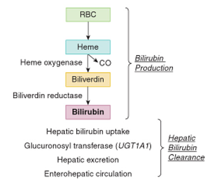
There is a reduction in the erythrocyte cycle in normal newborns (70 to 80 days in infants and 120 days in adults) increasing the amount of bilirubin in neonates. Hereditary hemolytic changes lead to increased RBC turnover and increased risk of hyperbilirubinemia. This causes hemolysis in newborns which is divided into several causes, namely first, damage to RBC metabolism resulting in G6PD and pyruvate kinase deficiency. The second is RBC membrane damage, of which congenital sporocytosis is an important factor. The third is impaired hemoglobin production where alpha thalassemia syndrome is the main cause in newborns. Fourth is an inherited immune hemolytic disease called medelian trait [6] (Figure 1). Schematic drawing of bilirubin production in neonates [7]. Bilirubin metabolism occurs in three phases, including the prehepatic, intrahepatic and posthepatic phases. Dysfunction in one or more of these phases can lead to jaundice [8].
biomedres-openaccess-journal-bjstr

Figure 1.

a. Prehepatic jaundice

The human body produces approximately 4 mg/kg of bilirubin per day from heme metabolism. Approximately 80% of heme is the result of erythrocyte catabolism, with the remaining 20% resulting from ineffective erythropoiesis and the breakdown of muscle myoglobin and cytochromes. Bilirub that is formed will be transported from the plasma to the liver to be conjugated and excreted.

b. Intrahepatic jaundice

Unconjugated bilirubin is both fat soluble and water insoluble, and therefore can easily cross the blood-brain barrier or cross the placenta. In hepatocytes, unconjugated bilirubin will be conjugated with sugar catalyzed by the glucuronosyltransferase enzyme and finally dissolved in bile.

In the intrahepatic phase, disorders associated with bilirubin can be divided into three subgroups [5,9]:

1. Excessive production of bilirubin

2. Disturbance in the conjugation process

3. Impaired excretory function

c. Posthepatic Jaundice

After dissolving in bile, bilirubin is transported through the bile duct and cystic duct for temporary storage in the gallbladder, or through the ampulla of Vater and into the duodenum. In the intestine, some bilirubin will be excreted in the feces, while the rest is metabolized by the normal intestinal flora into urobilinogen and will then be reabsorbed. Most of the urobilinogen will be filtered from the blood by the kidneys and excreted in the urine. A small amount of urobilinogen is absorbed in the intestine and excreted into the bile [8,9].

Jaundice can be classified into three main groups. This division is based on the presence of bilirubin found in serum [10]:

a. Unconjugated (indirect)

Albumin is insoluble in blood, therefore albumin is bound to blood. The free bilirubin is then combined with albumin and transported to the liver. In the liver there is an uptake mechanism, so that bilirubin is bound by liver cell membrane receptors and enters the liver. As soon as it is present in the liver cells, ligandin (Y protein), Z protein and other hepatic glutathione compounds occur which carry it to the hepatic endoplasmic reticulum, where conjugation occurs 4.

b. Conjugated (direct)

Conjugated bilirubin is formed in the rough and smooth ER, where it is conjugated with glucuronic acid. The result is water-soluble bilirubin (monoglucuronide and diglucuronide) a process that catalyzes glucoridation by uridine diphosphateglucuronosyltransferase, which occurs with the help of two isoenzymes. Excretion of bilirubin into bile is actively carried out by ATP-dependent transporters. Billirubin is transported into the stomach by the help of cells. Apolar glycuronide bacteria produce water-soluble unconjugated bilirubin, which can be converted to urobilinogen by reducing bacteria.

c. Albumin and bilirubin binding (indirect) [11,12].

Conclusion

Delta bilirubin is a small amount of conjugated illirubin. In serum irreversibly bound to albumin by covalent bonds. In laboratory tests to determine albumin levels, directly conjugated delta albumin is usually referred to as jaundice and is protracted, in which delta bilirubin levels rise proportionally and results jaundice.


For more Articles on: https://biomedres01.blogspot.com/

Friday, October 20, 2023

Scientific Results in Medical Education


Scientific Results in Medical Education

Introduction

The scientific results in Medical Education reveal the improvement in professional performance [1]. Ostgraduate education in the education system guarantees the permanent improvement of graduates’ college students Several training and development processes can concur in it, among which are: teaching-learning process (PEA), specialization, research, innovation, articulated in a relevant educational proposal [2]. Postgraduate education enables social development through continuous processes of creation, dissemination, transfers, adaptation and application of knowledge. Sustainable development through the training of professionals is closely linked with the practice that meets the demands of improvement with the in order to meet new challenges. The master’s degree is one of the ways of overcoming the academic postgraduate course that prioritizes the processes of productivity-focused learning. It aims to achieve a broad scientific culture in certain area of knowledge, greater capacity and development for teaching activity. in correspondence with the needs of production, services, economic, social, scientific, technological and culture of the country [1].

Health Technologies is a branch that contributes decisively to the solution of the problem health disease of Medical and Health Sciences. The technologist in the area in which he works has not received a continuous improvement that links this professional with the technological procedures, the technologies biomedical adjusted to the quality requirements that guarantee the epidemiological, clinical and social approach [3]. The results achieved in the area of technology are not at the desired level. They must provide a practical solution identified in the area in which they work. For this reason, the authors’ commitment to This publication is to define the scientific results of the training process of the master’s degrees in Information Technology Health.

Developing

The scientific results are the contributions that constitute products of the investigative activity. In them they have used scientific procedures and methods that allow solving problems of practice or theory. In general terms, world science is led by the great industrial powers such as: USA, Japan, UK and Germany. These countries are the largest producers of knowledge and scientific results [4]. In Latin America, they are dominated by large countries Brazil, Argentina and Mexico. The other countries in the region they barely contribute a small percentage of these results with international visibility. In general terms [5,6].

Latin America has Very Few Scientific Results in Relation to the World

In Cuba, the Science, Technology and Environment System is governed by the Ministry of Science, Technology and Environment (CITMA), in accordance with the methodological documents for the organization of science and technological innovation in the universities of the MES, 2017-2021. whose implementation consolidated scientific activity aimed at achieving scientific results linked to productivity and services [7,8]. The (CITMA), has worked, especially to fill the gap between knowledge and action. The scientific results in the health sector benefit population groups. The System of Sciences and Innovation Technology for Health (SCITS) is unique, it conceives the interdependence between teaching, services and research. As far as researchers have been able to systematize, the (CITMA) defines scientific results, it is the one that meets the requirements to be considered applicable. Features such as: novelty and contribution to the development of an activity, process or sphere of human knowledge that is supported by the technical commissions of experts constituted for this purpose [8].

Several authors have referred that scientific results are finished and measurable products. What do they contribute to from the material, human and available resources and the use of methods, techniques and procedures scientists. They achieve the specific objectives and contribute accordingly to the solution of the problem [6]. In the pedagogical area, the scientific result is the contribution to the solution of a research problem educational. Achieves from the available human resources the use of methods, techniques and procedures scientists to fulfill the objectives set and transform pedagogical practice or theory. The authors agree that the scientific results can constitute theoretical or practical contributions and should be meet certain requirements [9].

• Hat they are feasible: Real possibility of the use and of the resources that it requires; applicable clearly enough for implementation by others; generalizable, due to the condition of applicability and feasibility; that is valid: the result when it allows the achievement of the objectives for which it was conceived; clarity in the exposition, logic, coherence in the language used; rigor, the search for information, procedure and critical assessment; relevance, due to the social value and the needs to which it responds; novelty and originality. It reflects the creation of something that did not exist. Lazo M A, states that scientific results are “the product of a scientific activity, designed, planned and developed based on a social good, based on the use of scientific procedures, [10] directed to the search of solutions to the problems of the social historical practice”. Mastery is a scientific result “(...) the product of an activity in which they have been used scientific procedures, which allow us to offer a solution to something, is reflected in recommendations, descriptions, publications, containing scientific knowledge or material concrete production, or their combination and [9,10].

They solve a certain economic and social need. De Armas N, defined scientific results “(...) the contributions that constitute products of the activity investigation in which scientific procedures and methods have been used that allow to solve problems of practice or theory and that materialize in systems of knowledge about the essence of eleven object or its behavior in practice Travieso N, points out that the scientific results “(...) constitute products of the investigative activity in the which scientific procedures and methods have been used that allow solving problems of practice or [11] of theory and materialize in systems of knowledge about the essence of the object In this regard, they consider that in order to achieve a better understanding and application of scientific results, it must take into account that classification that divides them according to the aspect of reality that it transforms.

The theory or practice, although when the research is done the results are neither theoretical nor practical. They should be presented in a balanced way. The results express the achievements of the project, to what extent they reach the proposed objective. Must be concrete, measurable qualitatively and quantitatively. Related to indicators that verify it. I know verified through the presentation of publications of various types, presentations at scientific events, patents or registrations, prototypes, models, technologies, procedures or manuals. [11-14]. They also allow enriching, modifying or perfecting scientific theory. They provide knowledge about the object, the methods of science research, which can be classified into systems of knowledge and methodological on the other hand, the practical results have an instrumental character to transform the functioning of the object in reality making it more efficient, productive and viable.

They reach essential elements that characterize the effects and requirements that can be considered for determine the contributions in the practical theoretical plane of an investigation. It is used to provide pathways in the improvement of professional performance in the PEA. Show points of view, reflect on the eleven logical and methodological procedures underlying the construction of the proposal on the systematization carried out, the author identified the following regularities in the definitions of results scientists: finished and measurable products; scientific methods, techniques and procedures; problem solution; recommendations, descriptions; They solve certain economic and social needs. The application of results in the graduates of Health Technologies, for the improvement of the professional performance in the care areas where they provide services.

Allows to define the scientific result from the Sciences of Medical Education. The main author operationally defines scientific results from the Sciences of Medical Education and in particular the Health Technologies, product of the research process, which provides the solution of a scientific problem, which allows to identify, apply, process, make decisions for socialization and generalization of the results. It helps to transform reality.

Conclusion

The scientific results of the training process of the master’s degrees in Health Technology were defined, which enrich the epistemic bases of technologies as a branch of Medical Education, a science still in construction.


For more Articles on: https://biomedres01.blogspot.com/
Thursday, October 19, 2023

Carbon Dots as Antimicrobial and Antiviral Nanomaterials and Drug Delivery Applications


Carbon Dots as Antimicrobial and Antiviral Nanomaterials and Drug Delivery Applications

Introduction

Carbon Dots

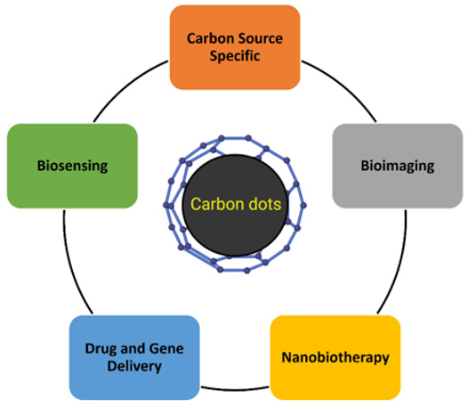
CDs a novel class of carbon nanomaterials discovered in 2004 have gained considerable attention due to certain specific characteristics: excellent photoluminescence, high quantum yield (QY), low toxicity, biocompatibility, small size, chemical stability, and inexpensive and easy synthesis [1,2]. The size of CDs is less than 10 nm, made of carbon, an element abundant and generally nontoxic. CDs are particularly attractive in many applications such as bioimaging, biosensing, and nanocarriers for drug delivery [3] (Figure 1). CDs are mainly classified according to their different formation mechanism, surface functional groups, and properties: carbon quantum dots (CQDs), graphene quantum dots (GQDs), carbon nanodots (CNDs), and carbonized polymer dots (CPDs). In addition, associations can be built among them by changing the carbonization degree and graphene layer (4).

Structure and Properties

Carbon dots have been characterized to analyze their physic properties and to understand the mechanisms associated with their properties. Common techniques are transmission electron microscopy (TEM) to obtain morphology, size, and agglomeration by demonstrating that they are very small of approximately <10 nm and quasi-spherical form, Fourier-transform infrared spectroscopy (FTIR) to identify specific functional groups, X-ray diffraction for the evaluation of the crystalline nature of CDs. It is also known that CDs are usually the product of the carbonization of organic precursors, mainly consisting of sp2 /sp3 -hybrid carbon or sp2 -domains embedded in amorphous carbon [5,6]. They also have several functional groups on their surface such as carbonyl, carboxylic and hydroxyl groups, which provide a rich part in oxygen and favor it to be soluble in water, in this way it can be useful in a wide variety of applications [7]. Carbon dots offer the flexibility to be able to manipulate both their structure and their properties depending on what type of carbon and organic molecules are made [8]. CDs have great properties such as
1) Optical absorption depending on the method of synthesis,
2) Excitation-dependent photoluminescence. The fact that the emission color of the CDs can be adjusted according to the excitation wavelength makes it very attractive for various applications.
3) Photo induced electron transfer,
4) Electrochemoluminescence,
5) Proton adsorption and
6) Low toxicity [3,9].

biomedres-openaccess-journal-bjstr

Figure 1: Biomedical applications of Carbon Dots.

Synthesis of Carbon Dots

According to the strategies, there are two types of synthesis ‘‘top-down’’ and ‘‘bottom-up’’ [10]. The top-down approaches include arc discharge, acidic exfoliation, laser ablation electrochemical oxidation, and microwave-assisted hydrothermal synthesis, where CDs formed from a larger carbon structure. This advantage is abundant raw materials, large-scale production, and simple operation [7]. The obtained CDs generally present graphite-like structures with weak fluorescence luminescence is a synthetic process and requires complex steps [11]. In the bottom-up synthesis, it has been used small molecules carbonized as precursors for example citric acid. This technique is efficient to produce fluorescent CDs. It has been produced under simple and mild conditions through microwave or hydrothermal reactions [12].

CDs Antimicrobial Activity

One of the biggest public health problems that fear is antibiotic resistance. This is due to the abuse of antibiotics, which has allowed new mechanisms of Resistance to be generated that is why new strategies are needed that are fast, easy to produce, affordable. These strategies include Carbon dots since it has been reported to have antimicrobial activity, due to its photosensitizer properties to produce oxidative stress by ROS and attack a wide range of microbes in the following ways: with physical or mechanical damage to the cell wall, disrupting the EPS matrix, causing biofilm splitting, inhibiting growth and even killing [13] Heteroatoms in carbon dots enhance the generation of ROS due to extra free electron incorporation in carbon dots. The functional characteristics of Carbon dots depend strictly on the precursors from which they are made, solvents that are used, as well as the functional groups of their surface. It has been shown that depending on the load of the carbon dot interacts or not with the bacteria. Something more interesting is that when preparing the CDs they can incorporate heteroatoms; they are called doped CDs, to make their activity more powerful [14]. Another strategy is to combine CDs with antimicrobial agents for example sodium hypochlorite (NaOCl), hydrogen peroxide (H2O2) to reach the maximum antimicrobial effect [15].

CDs Antiviral Activity

Viral diseases continue affecting millions of people around the world causing serious problems, many alternatives have been proposed to try to avoid them, and one of the strategies is CDs based on different carbon sources and synthesis methods, which can bind to viral proteins to stop multiplication or blocking the entry of the virus into the cell. The studies that exist are very recent but this activity has already been demonstrated with CDs from different methods and precursors, for example, curcumin carbon dots to prevent the entering and replication of coronavirus, benzocaine carbon dots to prevent the attachatchment of dengue, and zika virus, carbon dots to inhibit the binding of human norovirus, glyzhyrric acid carbon dots to inhibit the replication of herpes virus, boronic acid functionalized carbon dots to prevent entry step of human immunodeficiency virus 1, etc. The action of the CDs depends on the size, Surface, and load [16,17]. Lin et al., 2019 have suggested that C-dots can act at different levels of infection [18]. Actually, with the SARS-COV2 coronavirus pandemic in which there are no drugs to completely cure, CDs have been proposed as alternatives to stop the infection inside people’s bodies or detect it [16]. In a study conducted by Lai-Di Xu et al 2021, they used a new lateral flow immunochromatography technology in which they used red emission-enhanced CDs based-silica spheres as signals to detect SARS-COV2. In addition, the incorporation of the desired functional groups with QDs could effectively interact with the virus input receptors and affect genomic replication [19].

There are already some analyses that suggest that the cationic surface charges of CQDs interact with the negative RNA chain of the virus-producing reactive oxygen species within SARS-COV2. Also incorporating some specific functional groups to these CQDs would interact with the virus receptors and thus inhibit replication. One of the areas of opportunity to attack the SARS-COV2 virus is to look for alternatives to make CDs more efficient, perhaps using natural compounds to make them such as curcumin [20]. In general, we are facing a new field of research because although it has been shown that CDs have antiviral activity, many of these are in vitro and in vivo there are few so it is necessary to do more to understand the mechanism of action and know the real potential of these.

Conclusion

CDs have emerged as a new type of Nano carrier, breaking down barriers of production methodologies using carbon abundant raw materials and have begun to take an important place as nanomaterials with great potential for applications in the field of biomedicine. The antimicrobial and antiviral properties of CDs give them a promising future as new nano-drugs against resistant and emerging pathogens.


For more Articles on: https://biomedres01.blogspot.com/
Friday, October 13, 2023

A Case of Thoracic Disc Herniation Effectively Treated with Interventional Radiology Computed Tomography- Guided Percutaneous Endoscopic Radiofrequency Annuloplasty Using a Disc-Fx®


A Case of Thoracic Disc Herniation Effectively Treated with Interventional Radiology Computed Tomography- Guided Percutaneous Endoscopic Radiofrequency Annuloplasty Using a Disc-Fx®

Introduction

In 1972, Hijikata et al. established percutaneous discectomy (PD), which is a procedure aiming at symptom relief for disc herniation through a reduction in nerve root irritation associated with a decrease in intervertebral disc pressure [1,2]. Furthermore, herniation reduction is achieved through secondary cicatricial contraction of the intervertebral discs. PD has subsequently been modified to be a safer, noninvasive procedure, and has been applied in various forms, such as with the use of laser, including the Disc- Fx®. The Disc-Fx® facilitates nucleus pulposus transpiration or fiber ring thermocoagulation in addition to conventional nucleus pulposus extenteration using forceps (Hijikata’s procedure) [3-5]. In this study, we report a patient for whom Interventional Radiology- Computed Tomography (IVR-CT)-guided Percutaneous Endoscopic Radiofrequency Annuloplasty (PERA) was performed for the thoracic disc herniation to reduce the incidence of complications and improve treatment results, leading to a favorable outcome [6].

Case Report

The patient was a 34-year-old man (height: 167cm; weight: 74.4 kg), with a chief complaint of lightning pain in the right flank. He had no significant medical history. The patient consulted the Department of Anesthesia of another hospital with bilateral dorsal lower back pain, which had persisted for 1 year, and was treated with medication for 5 months. However, he stopped consulting the outpatient clinic due to symptom relief. 6 days before visiting our hospital, he again experienced lightning pain in the right flank appeared. 3 days before visiting our hospital, he consulted the Emergency Outpatient Unit of another hospital owing to exacerbation of the pain. Under a tentative diagnosis of ureteral calculi, an anti-inflammatory analgesic agent (diclofenac sodium suppository) was prescribed 2 days before visiting our hospital, he consulted the same hospital again, because there was no symptom relief. The presence of ureteral calculi was ruled out based on abdominal CT data. He was admitted with severe pain, and detailed examination and treatment were started. However, the presence of a disease in the field of internal medicine was ruled out. 1 day before visiting our hospital, he was discharged based on his wishes to consult another hospital. Although he consulted another local clinic, a definitive diagnosis could not be made. Next day, he consulted the outpatient clinic of our department. Physical examination on admission showed tenderness at the Th9 to 12 level.

biomedres-openaccess-journal-bjstr

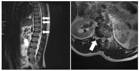
Figure 1:
a. MRI of the thoracic/lumbar vertebrae displayed multiple disc herniation involving the thoracic vertebrae to thoracolumbar junction (Th9/10, Th10/11, and Th12/L1). (arrows)
b. Coronal section of Th12/L1 showed a disc herniation (arrow).

biomedres-openaccess-journal-bjstr

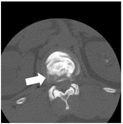
Figure 2: CT-discography led to a definitive diagnosis of lateral disc herniation at the Th12/L1 level (arrow).

In particular, there was marked pain at the Th12 level, and allodynia was noted at the site of the pain. Hematological analyses did not demonstrate any abnormalities. The Numerical Rating Scale (NRS) score was 10/10, and the Self-rating Depression Scale (SDS) score was 41. The EuroQol-5 Dimension (EQ-5D) score was -0.594. X-ray of the thoracic/lumbar vertebrae displayed osteophyte formation, leading to a diagnosis of thoracic spondylosis deformans. Magnetic Resonance Imaging (MRI) of the thoracic/ lumbar vertebrae displayed multiple disc herniation involving the thoracic vertebrae to thoracolumbar junction (Th9/10, Th10/11, and Th12/L1), suggesting thoracic disc herniation as an etiological factor for lightning pain of the right flank (Figure 1). Outpatient treatment was started. Intercostal nerve (right Th10, 11, and 12)/epidural (Th12/L1) blockade was performed, but there was no improvement. Blockade of the right Th12 nerve root was conducted under fluoroscopy, but the pain remained, with an NRS score of 5/10. Subsequently, during CT-discography, the patient felt irradiating pain which was consistent with the pain he felt constantly. CT-discography led to a definitive diagnosis of lateral disc herniation at the Th12/L1 level (Figure 2). Therefore, IVR-CTguided PERA with a Disc-Fx® that features a patented navigational device with bipolar system which was specifically designed to access and treat the diseased portion of the disc was scheduled.

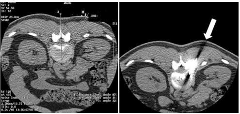
Under IVR-CT guidance, the point and route of insertion were determined, and a Disc-Fx® was inserted through an area 5 cm right to the spinous process at the Th12/L1 level to approach the intervertebral disc (Figure 3). Direct PD involving the herniated site was performed to treat the lateral herniation, and nucleus pulposus extenteration and thermocoagulation were conducted by DiscFx®. There were no complications, such as pneumothorax thoracic spinal nerve root injury and haemorrhage. After treatment, the NRS score was reduced to 1-2/10. Subsequently, follow-up was conducted by trigger point injection and drug therapy. 1 year after treatment, although there was a pain (NRS score was 2-3/10), there was no acute exacerbation of symptoms. The symptoms were calm, so it was decided to follow up at the clinic around the house, depending on the wishes of the patient.

biomedres-openaccess-journal-bjstr

Figure 3:
a. Under IVR-CT guidance, the point and /route of insertion were determined.
b. A Disc-Fx® was inserted through an area 5 cm right to the spinous process at the Th12/L1 level to directly treat the lateral herniation (arrow).

Discussion

PD is an intermediate treatment procedure between surgical and conservative therapies, which was proposed by Hijikata in 1972 [1]. It reduces pain by decreasing the intervertebral disc pressure and reducing stimulation-associated nerve root pressure, degeneration of the disc, and cicatricial contraction of the disc. Advantages of PD include low-level invasiveness, a low incidence of postoperative complications, and shortening of the admission period. Treatment as an outpatient is possible. As complications, persistent inflammation of the nerve root and intervertebral arthrosis associated with a decrease in the intervertebral space have been reported. Several studies indicated the efficacy of PD combined with epidural/nerve root/intervertebral joint blocking for persistent inflammation of the nerve root [7]. Recently, PD has been improved to become safer and less invasive, and various techniques, including the use of laser, have been applied. As one such technique, the Disc-Fx® was developed. This technique uses Trigger-Flex® and Surgi-Max® for nucleus pulposus transpiration by percutaneous high-frequency intervertebral disc decompression or fiber ring thermocoagulation in the process of intra-disc highfrequency thermocoagulation in addition to conventional nucleus pulposus extenteration with forceps (Hijikata’s procedure) [3,4,8]. In addition, this device facilitates the examination of the intra-disc nucleus pulposus and fiber ring under an endoscope.

Thoracic disc herniation is rare compared with cervical/lumbar disc herniation, accounting for 0.25% to 1.00% of all patients with disc herniation [8]. Symptoms of thoracic disc herniation vary, and include: dorsal chest pain, girdle pain, dysesthesia of the lower limbs (numbness, weakness), and myelopathy; therefore, an accurated diagnosis is difficult. In addition, treatment methods vary from conservative therapy to surgery. As this disease is rare, few studies have reported the results of treatment, and a consensus regarding treatment has not been reached. The present case was characterized by thoracic disc herniation and lateral herniation, which are rare. Therefore it was difficult to make a diagnosis. MRI of the thoracic/lumbar vertebrae, discography, and CT-discography led to a definitive diagnosis. The intervertebral foramens of the thoracic vertebrae are narrower than those of the lumbar vertebrae, and the lungs are present in the lateral regions; therefore, when performing PD for thoracic disc herniation, the risk of pneumothorax must be considered [9]. To reduce the risk of complications, we performed IVR-CT-guided PERA with a Disc-Fx®, although there are still few reports on this procedure. The point and route of insertion were determined under IVR-CT guidance, facilitating individualdifference- matched fine adjustment. Effective treatment could be administered while reducing the risk of pneumothorax by directly puncturing and extirpating the site of lateral herniation.

In addition, spinal canal operations and intervertebral joint drilling was not required, in contrast with surgical procedures; therefore, it was possible to reduce the risk of complications, such as spinal canal adhesion and thoracic vertebral instability. 1 year after treatment, although there was a pain (NRS score was 2-3/10), because the disc herniation other than lateral disc herniation at the Th12/L1 level remained. However, of course there was no acute exacerbation of symptoms in TH12/L1 level. In conclusion, although the long-term efficacy should be further investigated, the present case suggests that IVR-CT-guided PERA with a Disc-Fx® is useful for treating patients with thoracic disc herniation.


For more Articles on: https://biomedres01.blogspot.com/
Wednesday, October 11, 2023

First Twin Live-Birth Deliver After Oocyte Cryopreservation in a Follicular Lymphoma Patient


First Twin Live-Birth Deliver After Oocyte Cryopreservation in a Follicular Lymphoma Patient

Introduction

Many young patients diagnosed with lymphoma achieve durable remissions with current treatment strategies and become long-term survivors. However, chemo- and radiotherapy can determine a severe gonadotoxicity and compromise their reproductive potential [1]. Therefore, the demand for fertility preservation has greatly increased during the last decades and the matter is particularly challenging when female patients are concerned [2,3]. Oocyte freezing and thawing proved to be an efficient method for fertility preservation [4,5]. Nevertheless, small numbers of successful pregnancies and live births from oocyte cryopreservation have been reported in female cancer survivors, especially in patients with hematological malignancies [6-8]. This is the first report of a follicular lymphoma (FL) patient who carried out a healthy twin pregnancy through fertilization of the oocytes cryopreserved before chemotherapy.

Case Report

A 28-year-old nulliparous woman was diagnosed with non- Hodgkin FL, grade II, stage IIIA, in August 2014. She presented in good clinical conditions, asymptomatic, and objective examination only showed superficial lymphadenopathies, the largest ones measuring about 5 cm in her groin. Before starting hematologic chemo-immunotherapy, the young woman was referred to the fertility preservation specialists at our Institution and, after appropriate counselling regarding the available options, she decided to undergo oocyte cryopreservation. Since the patient’s hematologic condition did not contraindicate a moderate delay in the beginning of chemotherapy, controlled ovarian stimulation was performed: gonadotropins (follitropin α) associated with gonadotropin-releasing hormone agonist, namely leuprolide acetate. After 15 days of gonadotropin stimulation (the patient received a total dose of 1.575 IU), her estrogen level was 1.442 pg/ml and a transvaginal ultrasound showed 17 follicles ≥ 16 mm. Ovulation was triggered with α-chorionic gonadotropin (250 mcg). Fifteen oocytes were retrieved, 12 were cryopreserved through vitrification. No complications as ovarian hyperstimulation syndrome, bleeding or infection occurred after oocyte retrieval.

Ten days later, the patient started her hematologic treatment. She received 6 cycles of CHOP chemotherapy (cyclophosphamide, doxorubicin, vincristine and prednisone) associated with 8 administrations of rituximab, obtaining a complete response which was confirmed after 2 years of maintenance with rituximab.

In May 2015, after the end of chemotherapy, the patient’s menstrual cycle reappeared. She had fruitlessly tried to conceive for approximately 17 months when, in September 2018, she addressed the Infertility and In Vitro Fertilization Unit of our Hospital to research pregnancy with frozen oocytes. Endometrial preparation was performed with emiidrate estradiol patches, 300 mcg/day for 11 days. When the endometrial thickness reached 10 mm, 600 mg/ day of micronized progesterone were added. Four oocytes were thawed; two of them survived and were inseminated through intracytoplasmic sperm injection, resulting in normal fertilization with development of two embryos which were transferred into the uterus on day 2 after insemination. After 14 days the serum level of β-human chorionic gonadotropin was 537 IU/l. Two weeks later, transvaginal ultrasound examination revealed two intrauterine sacs with embryo heart activity. Gestational diabetes mellitus, treated with diet and exercise, was the only medical complication of pregnancy.

At week 36 of gestation, preterm prelabor rupture of membranes occurred. An urgent caesarean section was performed, which resulted in the birth of two healthy males weighing 2690 g and 2610 g, respectively. Uterine atony/postpartum hemorrhage occurred after caesarean section and was treated with Bakri balloon tamponade inserted within the uterine cavity. Patient gave written informed consent to publish her data.

Discussion

The increasingly prolonged survival of young lymphoma patients urges clinicians to focus on minimizing the long-term impact of antineoplastic treatment. An important issue is the potential toxicity of chemo- and radiotherapy on the gonadal function, which can lead young female patients to premature ovarian insufficiency and infertility [1]. Embryo cryopreservation is the most established option among assisted reproductive technologies, resulting in a pregnancy rate of approximately 30% [2]. More recently, oocyte cryopreservation has become a valid alternative for women who do not have a male partner and do not wish to resort to a sperm donor [3-5]. Overall, a small number of live births have been reported from fertilization of thawed oocytes in patients having received cancer therapies, most of them as single case reports or retrospective studies [6-8]. Some authors report that cancer patients tend to have a low ovarian reserve and an inadequate response to stimulation protocols, resulting in inferior numbers of oocytes retrieved when compared to age-matched non-oncologic women [9]. Others indicate that even the type of malignancy can influence the outcome of the procedure, with lymphoma patients showing lower levels of anti-mullerian hormone and needing higher doses of exogenous follicle-stimulating hormone compared to patients with solid tumors [10]. This aspect seems to be related to the high amounts of circulating inflammatory cytokines associated with this type of disease [10].

This topic is actually a matter of debate, since many other studies do not account for any significant difference in ovarian function among these categories of patients [11]. Another difference is that oocytes frozen for oncological reasons tend to face longer storage periods, due to the patient’s need for prolonged treatment; nevertheless, it was demonstrated that time of storage does not affect cryopreserved oocytes in terms of gene expression, provided the freezing is done correctly [12]. In fact, data show that no higher incidence of genetic aberrations or birth defects is associated with the procedure.

Conclusion

The case we described is, to our knowledge, the first twin live birth obtained through fertilization of frozen-thawed oocytes in a woman with a non-Hodgkin FL after first line treatment for the hematological malignancy. With this report, we aim to further stress the importance of oncofertility counseling in young women in need of antineoplastic therapy, in order to minimize the impact of gonadotoxic treatment on their future lives.


For more Articles on: https://biomedres01.blogspot.com/
Tuesday, October 10, 2023

Management of Penile Fracture with Urethral Injury in Haiti


Management of Penile Fracture with Urethral Injury in Haiti

Background

Penile fracture is a urological emergency requiring operative management [1,2]. Considered a rare and underreported traumatic injury [3-6], it has a reported incidence ranging from 0.29 to 1.36 per 100,000 people [1] and affects 1/175,000 men in the USA [6]. It is defined as a rupture of the tunica albuginea of the corpus cavernosum and occurs when the penis is erect [1,2,7] Erection causes thinning of the tunica, with its thickens reduced from 2 mm to 0.25-0.5 mm, and it becomes vulnerable to trauma [6]. The main cause of penile fracture is coital trauma, which represents 1/3 of all trauma to the penis. In very rare cases it is accompanied by rupture of the urethra which occurs in 1% to 38% of cases of penile fracture reported globally [1,2,8,9]. Some African studies revealed the main cause is masturbation [10]. Management of penile trauma has evolved over time. Initially, penile fractures were managed conservatively and non-operatively. However, due to the high complication rate observed, emergency surgical management has been the standard of care since 1980, which considerably reduces rates of long-term complications as erectile dysfunction [1,9,11-16]. To our knowledge this is the first described case from Haiti, we present a case of traumatic penile fracture and an associated urethral injury which presented to the emergency department at the State University Hospital of Haiti and managed operatively by the urology service.

Case Presentation

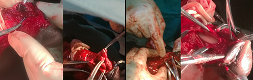
Our patient was a 49-year-old man with no known medical history. He presented to the emergency department with a six hours history of urethrorrhagia and difficulty of retracting the glans. He reported taking sexual stimulants and subsequently engaging in vigorous sexual intercourse during which he heard a sudden cracking noise at the penis after bumping the perineum of his female partner. He reported engaging in vigorous vaginal penetrative sex from the posterior position (i.e., doggy style). After the sound, he passed blood from the meatus, followed by pain and edema in the body of the penis. Given the increasing intensity of the pain and the persistent hemorrhage, the patient decided to come to the Urology Emergency Department. Initial clinical examination was remarkable for a leftward deformity of the penis, most markedly in the distal 1/3 of the organ, edema without apparent ecchymosis (Figure 1), and passage of blood via the urinary meatus. There was difficulty in retracting the glans and pain was most severe at the point of swelling. Examination of the scrotum and perineum was unremarkable. The diagnosis of penile corpora cavernosa fracture was made and the patient was consented and prepared for operative repair. The operation was initiated by the in ligature and section of frenulum artery followed by a circular incision was made through the balanopreputial groove to strip it down to the root (Figure 2). A tourniquet was placed for less than 30 minutes and exploration of the penis was initiated where clots were identified, hematoma was drained and wound was washed with 0.9% serum saline.

biomedres-openaccess-journal-bjstr

Figure 1: Description of the fractured penis with leftward deformation and edema of the penile shaft.

biomedres-openaccess-journal-bjstr

Figure 2: Circular incision and penile stripping.

Exploration of the penis revealed an injury in the corpora cavernosa on the right distal to the urethral meatus approximately 2.5 cm away. This was repaired with Vicryl 0, followed by a sealing test using 0.9% serum saline and removal of the tourniquet (Figure 3). After exploring the corpus cavernosum, we started the exploration of the urethra using a Nelaton catheter 16, which allowed us to identify and evaluate the anterior urethral injury which was approximately 3 cm in length and located at 5 cm from the urinary meatus. We proceeded to dissect the urethra circumferentially, then we proceeded to repair the urethral mucosal injury with (interrupted) Vicryl 4.0 suture over a 16 Fr. Foley catheter. A reassuring erection test with Nacl 0.9% was performed, and then we repaired the albuginea with (interrupted) Vicryl 2.0. Next, a second circular incision 5 cm (distal) from the first one was made, and we proceeded to repair the skin to the mucosa with a (interrupted) chromic 2.0, followed by the repair of the frenulum. At the conclusion of the procedure a sterile dressing was placed and the Foley catheter remained in place. The procedure which lasted approximately 2hr 45 min.

biomedres-openaccess-journal-bjstr

Figure 3: Circular incision and penile stripping.

Discussion

Penile fracture is the traumatic rupture of the albuginea attached to the corpus spongiosum [1,3,17]. This structure has a bi-layer of collagen, which provides a significant tensile force resisting the intracavernous pressure which can rise to1500 mm Hg. This tensile strength is essentially provided by the outer layer of the albuginea, the thickness of which decreases considerably during erection (2 mm to 0.2-1.5 mm) and thus weakens the corpora cavernosa [6,9]. Any situations or evens which elevate the intra-cavernous pressure above 1500 mm Hg are a risk factor for penile fracture due to the rupture of the albuginea and the corpus spongiosum [18]. These situations include the use of oral sexual stimulants and sex under psychological pressure, like extra-marital sex [2,6]. Our patient admitted to having used oral sexual stimulants prior sexual act. Diagnosis of penile fracture is essentially clinical [5,15,16]. The majority of cases involve a history of trauma, especially and more than a third of cases report coitus [1,2,19]. Other causes of trauma can lead to penile fractures, such as a fall from the top of the bed during sleep, direct blunt impact, and penetrating injuries such as that made by projectiles [1,2]. It is uncommon to see penile fracture with concurrent urethral rupture. The prevalence reported varies by region of the world. It is 3% in Asian and African countries, while it is around 38% in Western countries [6,8]. Furthermore, certain sexual positions favor the occurrence of penile fractures with or without rupture of the urethra.

Among these positions, the doggy style position and the position where the woman is on top are the most often described cases [14,20-22]. However, other positions can result in penile fractures as well. The most consistent clinical sign of penile fracture with urethral rupture is hematuria, followed by urethrorrhagia, dysuria, and urinary retention [5,18,23,24]. However, the absence of these signs does not exclude a penile fracture with urethral injury [13,18]. Furthermore, there are the classic signs of penile fracture such as pain, edema, ecchymosis, and the classic “eggplant” deformity [4,5,16,18]. Our patient presented with a similar picture; he had urethrorrhagia, edema, pain and an eggplant deformity. Imaging studies can be used in certain well-determined circumstances, among them sonography and cavernosography, retrograde urethrography, MRI, and cystourethroscopy [15]. These are not recommended in emergencies where the clinical exam is usually sufficient to make the diagnosis and to expedite emergent surgical management [12]. This was the case for our patient who had almost all the clinical signs, and thus we expedited his penile anesthetic block and operative repair. Since 1980, emergency surgery is recommended in all cases of penile fractures. The British Association of Urological Surgeons recommends surgery within the first 24 hours [14] in order to reduce the risk of longterm complications. Bozzini et al. observed a complication rate of 7.6% in patients operated before 24 hours and 68.7% in patients operated 24 hours after injury [12,14].

Our patient underwent the procedure approximately 12 hours after admission. The wound was approached by a coronal suture (Figure 2), the most commonly technique used, which gives better exposure, improved post-operative aesthetics, and facilitates dressing changes [2,9,18]. However, some studies have shown that this approach is associated with risks of infection and necrosis [9]. The rupture of the corpus cavernosum was partial as is the case in the majority of penile fractures [9,12] and measured 2.5 cm, while the urethral wound was mostly anterior and 3 cm in length. Studies have shown that urethral rupture is most common in bilateral fractures [12,14,19] and is often ventrolateral where the tunica albuginea is thinner [14]. Reports recommend the use of urethrography or retrograde cystography in cases of fracture with signs of urethral rupture [12,14,19]. In our case this was not available, so we have used a 16 Fr catheter a very common practice and highly recommended by some researchers and we proceeded to operative exploration with ultimate repair of the albuginea as recommended [4,19] with Vicryl 2.0. . Our patient has had a good clinical outcome. Post-operatively, he was followed for 2 days in the inpatient surgical ward. He received antibiotic coverage with ceftriaxone. After discharge on post-operative day, he was evaluated regularly at 3-day intervals with changes of the petroleum-based dressing and re-application of topical neomycin for the first 15 postoperative days. The patient was instructed not to wet the wound and to continue the neomycin application until complete healing. The foley was removed one month post-operatively, and the patient had satisfactory urinary function. Return to sexual intercourse was advised at 2 months post-operatively.

Conclusion

Any patients presenting with hematuria and/or urethrorrhagia in whom clinical history suggests penile fracture, an evaluation of urethral injury must be performed. The diagnosis is essentially clinical and requires surgical intervention within the first 24 hours after injury. After operative repair, return to sexual activity is recommended after 6-8 weeks. In developing country like Haiti, with limited resources like cystogram or retrograde urethrogram, clinical evaluation and surgical emergency is key to avoid long term complications.


For more Articles on: https://biomedres01.blogspot.com/

Antimalarial Aloe Compounds

  Antimalarial Aloe Compounds Introduction Among the most prevalent diseases caused by protozoan parasites, malaria is caused by parasites o...