Saturday, December 9, 2023

The Role of Micrornas in Osteoarthritis


The Role of Micrornas in Osteoarthritis

Osteoarthritis

Due to the increasing lifespan around the globe, musculoskeletal disorders like Osteoarthritis (OA) tend to have a higher prevalence comparing to the past decades. As of 2017, more than 300 million people had been diagnosed with osteoarthritis. Therefore, early diagnosis and treatment becomes a serious matter considering the impact in the quality of life for these patients [1]. Osteoarthritis is a musculoskeletal disorder characterized largely by pain, join disfunction, synovial effusion and swelling due to the degenerative destruction of the articular cartilage. Articular cartilage lacks the capacity of self-healing, due to the lack of vascularization [2]. During the degenerative process occurs an imbalance between anabolic factors and catabolic factors in the favor of the latter.

Several matrix-degrading enzymes alter the structure of the extracellular matrix (matrix metalloproteinases, disintegrin, etc.), targeting especially type-II collagen or aggrecans [3-5]. Among the risk factors we can mention aging, metabolic disorders, cartilage injury, obesity and mechanical stress exerted upon the cartilage [6]. These factors that trigger the disorder bring several small-scale alterations like genomic instability, epigenetic alterations, altered intercellular communication, telomere attrition, mitochondrial dysfunction, cellular senescence, stem cell exhaustion, etc. [7].

MicroRNA

The discovery of the first microRNA (miRNA), lin-4, in 1993 opened a new door for studying molecular biology [8]. Nowadays, MiRNA is considered a target for both diagnosis and treatment of OA by regulating the endogenous miRNA [4]. MiRNA are tiny, singlestranded, noncoding RNA molecules that regulate gene expression, having an average length of 22 nucleotides [9,10]. In most cases, miRNAs interact with the 3′ UTR (UnTranslated Region) of target mRNAs, suppressing the expression of the genes. In other cases, miRNAs interact with other regions, such as the 5′ UTR, coding sequence, and gene promoters. Also, miRNAs are involved in activation of gene expression under certain conditions, regulating cell cycle, apoptosis or differentiation [8,11,12]. There are several types of endogenous RNA molecules, like transfer RNA (tRNA), ribosomal RNA (rRNA), small nucleolar RNA (snoRNA), small interfering RNA (siRNA) and microRNA (miRNA) [9]. MicroRNAs can be identified in several subcellular structures: mitochondria, endoplasmic reticulum, P-bodies, nucleus, and nucleolus. Also, miRNAs can be found outside the cells via exosomes and detected in plasma and other bodily fluids [13]. Several miRNAs were identified in OA pathology, having an abnormal expression. The most common miRNAs are miR-9, miR-27, miR-34a, miR-101, miR-107, miR-140, miR-146a, miR-558, and miR-602 [14].

Mirna Detection Methods

The first miRNA isolation and detection methods used phenol-chloroform associated with RNA precipitation [15]. Nowadays, as the technology improved, many techniques were developed. Some authors divide these techniques in 2 categories: traditional techniques and modern techniques [16]. The most common techniques used are qRT-PCR, in-situ hybridisation, RNAsequencing, microarray, northern blot. qRT-PCR. It allows the instant detection and the quantification of genetic products generated during the repetitive PCR cycles [17]. RT-qPCR is a combination of three steps: RNA to cDNA conversion using reversetranscriptase (RT); PCR-based amplification of the cDNA; real-time detection and quantification of cDNA [18]. In-situ hybridisation. It can localize genetic material in a sample by hybridizing and labeling a complementary strand of RNA or DNA. Using the labeled strand, a certain sequence of nucleic acid can be identified. The detection can be performed using isotopic or nonisotopic methods [19,20]. RNAsequencing. It can reveal the entire structure of a transcriptome using high-throughput sequencing methods.

It can also provide analysis for other aspects like gene expression, translatome, alternatively spliced genes, etc [21,22]. Microarray. It can analyze simultaneously the expression of thousands of genetic sequences in a single experiment. The sequences are arranged in a row-column array on a glass slide known as “chip”, facilitating their identification [23,24]. Northern blot. No special equipment is necessary. It uses the following steps: an agarose gel electrophoresis is performed in order to separate RNA sample (separation is performed according to the size of the sequence); it is transferred to a nylon membrane (preserving the separation in the gel and keeping the same arrangement), fixed and labeled by marking it with an isotope. A wash is performed, removing the unnecessary marked probes. The analyze can be performed using autoradiography or other techniques [16,25]. Despite the differences between these techniques, the results depend firstly on the quality of the sample. Secondly, not all detection methods cand isolate the miRNAs equally. Different extraction techniques isolate different RNAs, depending on the length of the molecules, their concentration in the sample and the sequence differences between miRNAs [11,26]. As a downside, most of the detection methods require long processing time, laborious techniques, and provide many false-positive results. Thereby, at the moment there is no universal detection method for miRNAs [16].

Epigenetics and MiRNA

Epigenetics affects OA in two major ways. First, the development of joints and bones is regulated via epigenetic mechanisms. Any change in these processes modify the risk of developing OA at some point in life by changing the joint shape, the extracellular matrix composition and/or the responsiveness of joint cells to cytokines and growth factors. Second, epigenetics processes can be triggered by external factors, such as articular traumatic injuries or metabolic disorders [27] Inflammation. The inflammatory process induces early alterations in cartilage structures way before the appearance of radiographic signs in OA. Micro fissures in articular cartilage and ECM catabolic products have been discovered in the synovial fluid in the early stages of OA [28]. MiRNA expression is determined by proinflammatory cytokines that lead to activation of the target genes that induce OA progression. Also, miRNAs are linked to modulation of proinflammatory cytokine expression, such as TNF-ɑ, IL-1β, IL-6 [2,12]. Also, there are high CRP serum levels that correlate with the histological alterations in the synovial inflammatory site.

Due to high activity of IL-1β and TNF-α, there is an overexpression of other critical inflammatory and chrondrolytic mediators, including MMP-1, MMP-9, MMP-13, NO, PGE2, and IL-6. IL-6 acts as B-cell and T-cell activator, but also as a regulator for the recruitments of other inflammatory cells. IL-8 has a synergic effect along IL-6, recruiting and activating neutrophils [28-30]. Proliferation. In normal synovial membrane, the thickness is about 1-3 cell layers and a low level of inflammatory cells in the synovial fluid. In OA, however, the synovial reaction consists in hyperplasia associated with a high degree of inflammatory cells, mainly macrophages, B and T cells. During the early stages of OA, due to the increased cellular activity at the site of the articular cartilage, chondrocytes tend to form clusters consisting of 50 or more cells. On the other hand, simultaneously with the chondrocyte proliferation, ECM tends to decrease, having a reduced level of glycosaminoglycans in its composition compared with a normal ECM, resulting in degradation of intraarticular homeostasis and opening a door for further destruction mechanisms.

During the late stages of OA, the cartilage structure consists of hypocellularity due to a high level of chondrocyte apoptosis and lacunar emptying [28,31]. IL-6 and IL-8 are the main proliferationinductors involved in OA. Recent experiments revealed that miR-373 acts as a downregulatory for expression of IL-6 and IL-8 by inhibiting a specific receptor called P2X7R. In OA, the plasmatic level of miR-373 is lower when compared with the non- OA plasmatic level samples [32]. Also, the expression of MiR-27b and MMP-13 activity is inversely proportional, miR-27b acting as a negative regulator. Mir-488 inhibits MMP-13 via Zinc-transporter 8 (ZIP-8), thus enhancing both chondrocyte differentiation and cartilage development. In OA, its expression is strongly diminished [10,33]. ECM degradation. ECM of the cartilage is composed of proteoglycans such as aggrecan (the main component), decorin, lumican, and biglycan; adhesive glycoproteins (fibronectin); collagens, mainly type II and in smaller proportions type VI, IX and XI collagens. These glycosaminoglycans (GAGs) have an absorptive function, increasing water concentration into cartilage ECM, thus enhancing compression resistance.

Type II collagen provides cartilage mechanical resistance to tension due to the fibrillar structure. Proteinases released in the articular cartilage in OA play a crucial role in degradation of ECM by targeting mainly the aggrecans and the collagen, damaging the two main components of the ECM. The destruction of these structures is mediated mostly by collagenases (MMP-1, MMP-8, MMP-13) and aggrecanases (ADAMTs) mainly via IL-1β, TNFα [34-36]. There are several other factors that contribute ECM breakdown such as Gc-globulin, α1-microglobulin, and α2-macroglobulin, but also VEGF. Also, IL-1β acts as a downregulatory in production of type II and type IX collagen, thus inhibiting ECM production [28]. Several miRNAs play a role in cartilage protection by modulating ADAMTS-5 expression, resulting in preservation of extracellular matrix. One of the first “protective” miRNA discovered is miR-140 that contribute to articular cartilage and normal enchondral bone development. In OA, miR-140 is downregulated compared with normal cartilage [2,37,38]. Apoptosis. Inflammatory cells migrate to the site of the injury in order to initiate tissue repair. After the reparation process is finished and the cells completed their task, they are eliminated via programmed cell death or apoptosis in order to prevent excessive inflammation [31].

Predominant factors that regulate apoptosis include enzymes, genes and proteins such as p53, Fas receptor, BCl-2 and Bax, cytochrome C, caspases, protein kinases regulated via extracellular signaling. The first stage in the apoptotic process is represented by the genetic control that decides, according to a stimulus, if the apoptosis should be initiated. It is regulated by two genes, BCl-2 and p53. The second stage is represented by the morphological alterations of apoptosis regulated by caspases [39]. Chondrocyte apoptosis may be linked directly to the destruction of the ECM. Most cells attach to the neighboring cells or ECM, creating junctions and ensuring the necessary nutrient intake in order to sustain their activity. Disruption of ECM may affect chondrocyte survival, inducing premature apoptosis.

In vitro experiments using enzymatic treatment via high purity collagenase in order to cleave type II collagen proved that the degradation of collagen induced chondrocyte apoptosis [31]. IL-1β modifies the expression of collagen gene Col2a1, having a downregulation effect and also having an upregulation effect over inducible nitric oxide synthase (iNOS). This enzyme has a chondrolytic effect over the cartilage, promoting apoptosis in chondrocytes. Along IL-1β, miR-101 is also an important factor that promotes chondrocyte ECM degradation. MiR-34a can decrease the IL-1β-induced effects [10,40]. Cartilage injury initiated by mechanical pressure leads to a high expression of miR-146a, having an apoptotic effect in human chondrocytes by inhibiting of Smad4. Its expression decreases with the OA progression [31,41]. Autophagy. In order to maintain homeostasis, living organisms tend to trigger mechanisms involved in cellular turnover. Autophagy represents the process regulated by several autophagyrelated genes like Beclin-1 and Light Chain 3 (LC3) that dismantle damaged or unnecessary cells and their components prior to their removal [42].

The expression of these genes is directly proportional to the metabolic activity of the involved tissue [43]. Depending on the region of the cartilage, the expression of the autophagy-related proteins differs. Thus, in the superficial layer, cells display a higher expression of proteins like BECN1, ATG5, and MAP1LC3. The cells located in the deep layer have a lower expression of MAP1LC3. Also, their expression decreases with aging. Once the autophagic process is disrupted due to the reduction of these proteins, apoptotic activity increases [44]. In OA, miR-107 acts as an autophagyinductor, having a protective effect on chondrocytes and reducing ECM degradation [33,45].

Potential Therapeutic Targets

Current management of OA involves pain-ameliorating medication and joint replacement surgery. None of these treatments adress to the underlying cause of OA. Due to this fact, there is a need for a Disease-Modifying-OsteoArthritis-Drug (DMOAD), a treatment that not only reduces the symptoms, but also interfere with the pathophysiology of OA, stopping the progression and at least prevent further joint degradation [46,47]. Currently, miRNAs represent a therapeutic target in the management of OA. So far, the clinical success of miRNA-based treatment is not satisfying. Due to the very unstable nature of miRNA, it has a short half-life, it is unstable in vivo and interferes with the disruption and saturation of endogenous RNA. Also, because of the lack of vascularization, miRNA hardly reaches chondrocytes in order to exert the expected effect. Therefore, various techniques must be developed to acquire a better miRNA stability and also a proper drug-delivery technique [48]. There are early studies involving miR-140 treatment in rats where the miRNA was administered intraarticular via exosome. It was observed that the chondrocyte numbers and cartilage thickness were greater after the miR-140 treatment and a reduction in MMP- 13 and ADAMTS-5 expression, thus reducing the ECM degradation and reducing OA progression [49,50].

Conclusion

MicroRNAs have a diversity of biological functions, being involved in a wide range of pathological processes. Also, they regulate gene expressions, activating or inhibiting gene expression. In the pathological mechanism of osteoarthritis, miRNAs have a regulatory role, so they are considered to be potential targets for diagnosis and treatment of OA. Present research directions are effectively promoting miRNA study, due to the increased potential of becoming diagnosis and treatment biomarkers of OA.


For more Articles on: https://biomedres01.blogspot.com/

Friday, December 8, 2023

Time to Exceed the Physical World to Reach the Quantum Dimension


Time to Exceed the Physical World to Reach the Quantum Dimension

Introduction

Dark Space and Universe Energy

Dark dominates the universe. The source of light is also darkness. Accordingly, “dark light” is the central concept of the universe. Because of darkness, galaxies are held together, and that’s how gravity is provided. Darkness is found throughout the universe. There is also space in every dimension of the universe. This gap allows communication between objects. Thus, in the dark space, there’s an electrical current, which is a magnetic attraction area. This magnetic field moves mass. The magnetic field created by the dark space is the magnetic field in which the movement and flow of objects in the nucleus of the mass in the universe occurs. Thus, galaxies, planets, and objects in the universe don’t all rotate in the same direction. Some stars have very little or little light because of the magnetic field they have. But not all parts of the universe have the same levels of a magnetic field. The same is true for the human body. Just like the Earth, planets have the magnetic field of space. Where there is magnetic field, there is life. This is because the magnetic field, the planet, has an atmosphere in its own right and in accordance with its conditions. The magnetic field in space is not the same everywhere. Thus, the effect of this magnetic field is different. Scott Kelly was on the International Space Station for about a year. When he returned to Earth in 2016, he was noted to have grown five centimeters. Kelly says it’s easier to get used to living in space than to living in the world. Accordingly, human body cells can adjust to any environment.

The fact that even telomers have this feature demonstrates that all cells and organs in the human body have a quantum size. Thus, the human body is three-dimensional with all of its cells and organs. The formation of the universe and the human magnetic field takes place in the space environment. The magnetic field generates energy. The energy in the universe is fed by this dark space. Accordingly, this “Dark cosmic light” is the main energy that feeds the universe and humans. Every creature in the universe has another magnetic field, except for its own magnetic field, which is made by a dark space. Just as an atom has a magnetic field, the Earth and the galaxies have their own magnetic fields. A magnetic field is one of the forces that play a role in the transformation of energy in the universe from one form to another. Because everything in the universe has its own magnetic field, we perceive the universe as material and see it as the universe. We can only see part of the universe. We cannot see the rest of the universe. The reason for this is that the dark energy causes the universe to accelerate into expansion. The gravitational effect of dark energy drives this. It alienates objects and accelerates the expansion of the universe. Where a magnetic field is generated, there is magnetization. In the universe, energy is drawn into space. Thus, the energy is in the voids. The dark energy that occurs in space accelerates the expansion of the universe. Likewise, human gaps maintain energy and promote human development in the human body.

As a matter of fact, a fetus is in the abdomen of the womb and continues to grow and develop in the space. The organs in the human body have their own voids, which produce magnetic fields. The lobes in the brain, some organs of the human body such as the lungs, kidneys, eyes, and ears are dual to produce magnetic fields in the voids. In this case, these organs form magnetization because there are molecules in voids that collect light. These light-containing molecules change shape with light. Then they recharge in the dark. If we take the human eye out of the socket and replace it with a new artificial eye, the light coming into the eyelid will stimulate the cells inside, send a signal, and start to see again. On the other hand, if we cut the nerve away from the brain’s sensory field, the brain will continue to function. Neil Harbisson was naturally colorblind and could “hear” the colors with an aerial attached to his head. Likewise, people without both hemispheres gain the functions of the brain. Humans have specialized tissues where chemicals in the air (in space) that can interact with neurons. Neurons in the human body have multiple processes. For example, in parts of the brain (occipital) that are associated with vision, the lobe also carries hearing and touch information. Also, areas outside the visual cortex allow us to see some neurons. Therefore, if neurons in the brain enable vision, neurons in the heart and other organs perform the vision. The act of seeing is not just about the eye.

The human genome has genes that enable us to see. There are two types of vision genes. One is a gene that’s formed in cavities, and the other is a gene that’s specialized in the air to enable the chemical to interact with the neurons to see. The phenomenon of human vision occurs when light enters into an eye space rather than when it is transmitted through the eyes. Accordingly, the light, which is conscious through the development of magnetic field, undergoes energy transformation. When conscious light interacts with an intensive magnetic field, changes occur within nerves and spaces. Every neuron in the human brain has a different photon. Light is therefore conscious and has the same effect on physical, chemical objects and gases in the human body, but some chemicals and gases create intensive magnetic fields. Cells use the magnetic field that forms in space.

Dark Space and Time

Mobility occurring in the universe is related to the direction of movement of the objects. The quantum-sized notion of time creates time based on the magnetic environment. Thus, the time of planets is different. The quantum mechanism walks with time. But the quantum dimensional time is different from the time in the universe. Therefore, as the size of the universe jumps, time changes according to these dimensions. Time is associated with motion in the universe. There are proteins that are produced by movement in the human body. Like the universe, the human body has a space in which time passes backwards.

Dark Cosmic Light

It’s light or optical light that we see for things. But there are forms of light that are not visible around us like ultraviolet light. According to physicists, light is the name of a physical being. There are many light types such as etheric, infra, ultraviolet, electra, gamma, and nucleon lights. Light is mainly used for 12-20 unit TV series, but is also longer series such as black light. But even though it’s said that the eye can’t detect such light, the eye can actually detect them. It is just that the enzymes and proteins to ensure this perception do not become activated and the eye cannot perceive these lights because perceiving light is dimensional work. The array-length difference in the light also makes its energy different. If it weren’t for this range difference, the messages from the universe and other things would only tell you their fractals, but not their properties. Therefore, the messages the eye will send to living creatures should not be based on certain light waves, like an optic spectral analyser that sees the detection center, but on the energy difference. The transfer of consciousness starts in the space that forms the universe and in the Dark Cosmic Light Alemdar [1]. Dark cosmic light is linked to all dimensions in the universe. If man transfers energy from cosmic light in the universe, it raises its own consciousness.

Space connections in the human body are essential in order for the body to function properly due to their role in electrical connection. Anatomic gaps exist in the human body. Consciousness transfer is also performed during sleep by discovering body cavities. This space is where the main source of energy is generated that triggers everything in man and the universe, which is a great source of energy where everything moves. Thus, what we call the void, which exists in the universe and in man, is full of energy. There are transitions in the dark space. There are magnetic pathways that happen in those spaces in the universe and in humans. Consciousness arises in the cavities of the human body. Spaces in the human body are the most suitable places for observing dark energy. Both dark energy and physical matter are found in voids. Therefore, dark energy can be detected.

Consciousness

There are ninety-nine types of consciousness. Within these ninety-nine consciousness, there are special consciousness. There are 360 aspects of consciousness. One in every way helps with information from the magnetic fields that form in the human body, which is to say, the way the mind activates. That is, every person has a different level of consciousness. In this direction, every person has a frequency level. Although this level of awareness can be raised, not every person can attain the same level of awareness. Ninetynine conscious species have different frequencies. Humans are only aware of the consciousness of visible light. However, a human lives in different frequency sizes. With different frequencies, there are ninety-nine dimensions formed by ninety-nine consciousness in the universe. The cells in the human body also have a special consciousness. These cells have special time zones where the consciousness of these cells activate. Therefore, each nerve in the human body is sensitive to photons of different colors.

Sleep as a Type of Consciousness

Sleep is connected to universal energy. Therefore, consciousness transfer takes place during sleep. Awareness is raised in the body cavities, and then energy is transferred between the universe and this vacuum. Human memory is accumulated in body cavities. Different parts of the brain have different tasks. These zones are responsible for language, memory, consciousness and intelligence. Emotions such as memory, thinking and feeling are created in spaces Reddy, et al. [2]. While the responsibility for these tasks initially burden the brain, in fact all organs of the body assume these functions. The major consideration here is that brain organs tend to think, act, and receive signals, which are actually developed in gaps in the human body. As such, what do these parts of the brain actually do when you consider people without brains or other living things? In fact, the entire body is in motion and all body cells are constantly renewed, developed, and divided. Neurons do not only belong to the brain, but also exist in other organs of the body such as the heart. All parts of the brain are like a distribution site. Since everything happens in space, these parts of the brain are dissipating energy. There are pathways in the space and light is emitted from these pathways to the whole body in different ways Alemdar [1]. Accordingly, emotion, thought, and automatic movements of people are caused by the spread of consciousness that is formed in different sizes than the pathways of the human body.

The magnetic field in the brain transforms light into a form other than the initial form Kletetschka [3]. The sensing and talking points in the brain are distribution centers. There are proteins inside the brain that produce magnetic waves. The effect of the magnetic field allows the neurons to produce signals. During the fourth week of fetal formation, the fastest developing cells are the nerve cells, the neurons. This is the longest-lasting group of cells. These cells survive without rejuvenation. At the end of the fourth week, neurons leave the spine. Neurons know what to do and where to go on this journey and they never miss the way. How do neurons find their direction without any mistakes? The answer is cells that have a magnetic field in the human body and the magnetism that occurs in that area. This creates a special consciousness in cells. A certain amount of sleep is necessary to maintain consciousness. In fact, humans move to the consciousness phase when they sleep and become open to the magnetic fields of the universe. Thus, a quantum-sized human can initiate the transfer of consciousness during sleep. The person is dismissed from the outside world while asleep. When asleep, neurons gradually calm down. Electrical wave activity begins with the magnetic field in space and light consciousness occurs within the human body. When a person has little sleep, an insufficient amount of magnetic fields and energy is created in the brain or body cavity during energy transfer.

In this case, the human body begins to produce slow waves. This can affect human consciousness and, linked to memory, causes the development of harmful proteins. Sleep must be at night. Because at night, the pressure in the brain goes down. The human being is linked to another dimension in the REM stage Rué-Queralt, et al. [4]. In the moment of sleep, awareness is transferred through the tunnel connections I call “human wormholes,” which transfer consciousness outside of time and space. According to this, a dream is not a realm of fantasy. Man is connected to two lives and has a two-way body. These are the state of sleep with alertness. One feature of paradoxical sleep, called REM, are the signals that are propagated by the brain during this sleep phase. Consciousness transfer is shaped by the individual’s level of consciousness. When consciousness transfer begins during REM sleep, the brain’s emotional and memory aspects are activated first Rasch [5]. This is related to the transfer of consciousness into the brain cavity in REM sleep, and the arrival of inspiration that has led to many inventions during that time Hobson, et al. [6]. Paul McCartney, the Beatles’ star in the world’s most famous band, acquired some of his songs during this phase. Dmitri Mendeleyev, the inventor of the periodic table, also dreamed about it. Examples of this are numerous.

People who sleep less are more likely to develop sleep disorders such as sleep apnea, elevated blood pressure, heart attack or stroke. Non-sleeping people have a toxic sticky protein called beta-amyloid in their brains. Insomnia causes more beta-amyloid accumulation in the brain. There are a number of systems still unexplored in the human brain. These systems serve as energy distribution centers in the brain. Beta-amyloid is not only present in the brain, but also in other organs of the body. For example, beta-amyloid is located around liver and biliary ducts Babu [7]. Adequate sleep helps remove all the dangerous toxins accumulated in the brain, including betaamyloid. However, REM also appears to increase blood circulation and beta amyloid accumulation in the brain fluid. It turns out that this particular protein, when it accumulates in the brain regions, attacks and damages these areas. Lack of REM sleep reduces the capacity to clean out beta-amyloid. The information obtained by the brain is linked to each other during the transfer of consciousness through sleep to the brain cavity. People who do not sleep or do not draw enough energy during transfer from dark light at night damage their body organs and start to produce harmful proteins. At night, consciousness transfer occurs. DNA repair occurs overnight as well Zada, et al. [8]. Many things are being rebuilt during the night. We use another energy at night, and another energy during the day.

The universe, too, is fed by darkness. During the conscious transfer in the visible and invisible spectrum, there is a cycle of alertness and sleepiness. Magnetic bacteria, which may be the main source of magnetic sensors in humans, are synthesized in the intestine and concentrated in certain parts of the brain. Accordingly, the bacteria that produces magnetite in the human body allows the creation of dreams in the brain. Toxic gases also produce negative magnetic fields. Magnetic fields come into contact with each other through sleep-vigilance and interaction made as a result of day and night. During the transfer of dark energy from space at night, during REM sleep, acetylcholine and similar molecules are doubled, creating new bonds between it and the neurons. The magnetic fields that occur during REM, i.e., transfer of consciousness, are communicated with the neurons Pace-Schott [9]. This magnetic field in space, and the neurons, just like a magnet, pull one another. Since the transfer of consciousness takes place at night, this situation necessitates sleep. A heavenly body that rises to the earth from the dark at night has light and energy, and the process of transferring the body from the celestial body to the human being begins with the light/energy that comes at night. Humans are part of the energy that occurs within the dark space. The whole body nervous system consists of neuron cells, which carry electric and chemical impellers.

If you want to measure impellers of the nervous system, some are going at 200 miles an hour, some are going at 2 miles an hour. How is it that the brain and other parts of the body can coordinate the nervous system and our sensitive movements when these impellers travel at different speeds as predicted? Now this is a problem that needs to be solved. Here, this system needs a gap that explains how to coordinate. A review of nervous system activity reveals that it starts to fire simultaneous vibrations and pulses across different parts of the brain. When consciousness functions, these pulses start firing mainly coherently. Scientists, too, look at how all different parts of the brain in the same focus can quickly co-ordinate, and that compliance in firing is faster than the physical capabilities of cells communicating from one area to another. Therefore, essentially, these results clearly show that the brain communicates at a higher level than the physical transmission of nerves. And because you’re tuned to the energy in this body cavity, you’re constantly in contact with this information band. And the memory that is communicating in this information band is in the body cavities, including the brain. Because every cell has its own memory. Memory does not just belong to the brain. Subatomic particles extract energy in voids. The energy between the subatomic particles is formed in space. There is a large amount of energy in this gap. The energy in this space continues the invisible energy around us. This vacuum contains energy, or “consciousness information.”

This consciousness in the gap is always accessible, which allows a person to constantly draw information. Backster’s research on the subject is striking Backster [10]. He took DNA samples from subjects, put them in an isolated lab about 300 kilometers away, and urged them to change the minds of the subjects they belong to, and to think of bad events, to fear. The DNA in the lab completely changed and affected the subject’s DNA, 300 kilometers away. In my opinion, the occurrence of this effect is due to the aforementioned gap. There is no loss of information and time in the space where information is transferred. The brain makes up approximately two percent of the total body weight. A considerable amount of energy is used to support brain activity. Today’s research attempts to combine the human brain with artificial intelligence. Their work combines artificial intelligence and the human brain, and works on implant devices that connect a computer directly to the brain. These chips/implants should be placed in the cavities inside the human brain. This is the only way to succeed.

Synapses are the areas in the space where neurons transmit neurons to each other. Cells are often separated in an incredibly subtle space called the synaptic space. The brain communicates with its surroundings through sensory organs that react to various stimuli such as light, sound waves and pressure. Sleep-wake cycles are controlled by neuro transmitters that act on different areas of the brain to initiate and wake sleep. If a person does not get enough sleep, the ability to think and remember clearly and rapidly decreases.

The person’s frequency is high during sleep Whitmore [11]. This is because the consciousness during sleep is in communication with the universal consciousness. It thus dominates the nervous system and, through it, the organism. People are usually on the move during the day. At night, the opposite is partially inactive. Centers that appear inactive and passive to the environment during sleep achieve important tasks inside. The effects to the brain increase the movement of the molecules that make up the centers of the brain. The human body is made up of enzymes and cells that work day workday and night in a day and night cycle. Some cells work in the dark: some need to work during the day. That’s why one sleeps and is awake. Sleep is a state of awareness. Studies on sleep deficiency have shown that people who regularly get little sleep have less concentration and make more mistakes. Weakening of the immune system, changing hormone levels, obesity, depression, anxiety, diabetes and heart disease are the main causes of low sleep. If you do not comply with your needs and draw energy from others, your body may experience inconsistent behavior. This causes some organs to become destabilizing in various cases and may result in harmful proteins. This leads to a number of diseases and toxic gases. Gases are also affected by magnetic fields. When vibration deteriorates in the human body’s organs, it begins to break down and scatter, which means that the consciousness that dominates these cells has lost interest in the body.

Dark Matter

It’s not dark matter that’s effective in the universe, but dark energy. There are various forms of light coming around us from the dark space, such as rays of photons. Humans, too, have special proteins and cells specialized in detecting this light. The energy or photon transferred in space communicates with the photoreceptors as a result of the magnetic field. It’s recognizing the signals that the photoreceptors send into the spaces as human images. Each of these receptors is set to different types of consciousness, light. Some of these cells work during the day, and some work during the night. The cells, which work during the day and night, are sensitive to different sizes and types of light. For this reason, the night, rather than the day, shifts to quantum size with human organs and cells. The eyelids and eyelids are arranged according to night and day. The orbicularis oculi muscle in the human eye, like the eyelids and Saturn rings, defines the direction of the magnetic stripes in the eye. The visible light breaks the photon through the rings around this muscle and adjusts the intensity of the photons reaching the eyes by protecting it. This muscle is for the blink of an eye. What is seen during the day, and what happens in the night is different. Indeed, one of the nerves in the eye is optical vision. The other one is for quantum vision. Even when the eye muscle is closed, it produces an incredible magnetic force.

A person has two distinct dimensions of two human status. The physical human condition is more about day. The second dimension of the human being is about night. The physical size of the human being slows down with sleep at night, decreasing the material side and strengthening the quantum dimension. The size, which must be fed by dark energy, is fully active during the night. All the cells of a person work at full capacity during the day. At night, the material size of human metabolism and many of its physical activities are suppressed. According to this, the energy between day and night is different. Energy flows into the human body at night, and during the day it is adjusted by the auxiliary organs and muscles. The survival of consciousness in man requires movement, change and revelation. That’s why so far the puzzle of matter and consciousness has not been solved. So, the stuff that’s called matter is created by the motion and the magnetic fields in the universe. Global consciousness cooperates with dark energy. Universal consciousness reaches the human body through wormholes in humans. This state of consciousness in people manifests as vibration and energy. That is, human consciousness depends on the action of consciousness because of the energy that it gets from magnetic fields. So everything seems to be a vibrating matter to man.

The body is made up of cells. Each cell has its own unique frequency Jafari [12]. The higher the frequency of a cell, the greater the ability to use that cell at a quantum size. The cells with the highest frequencies in the human body are brain cells. The parts of the brain that have the highest molecular frequency are magnetic fields. The increasing frequency here increases the activity in the human brain and other organs. The expansion of consciousness depends on frequency increase. The increase in frequency depends on the energy from dark energy. The brain has about 90 to 100 centers. Energy is stored in the brain and other gaps. The physical and spiritual development of man means that cells are constantly in motion and evolve. The energy from dark space is stored in body cavities, where the magnetic field evolves into an electrical signal, and a chemical signal. There are magnetic fields in the universe, solar system and planets. There are magnetic fields in the human body. The universal consciousness that dominates the entire universe arises in these magnetic fields of the human body. Unconsciousness on these magnetic fields, which originate in voids, continues to exist. The consciousness undergoes various changes as dark energy is linked to consciousness and a magnetic field is developed. A magnetic field is not energy. The magnetic fields of light, which we call consciousness, are transformed in the brain and body cavities.

If an intensive magnetic field is formed in the body, the conscious light is changed. Gases, chemicals and certain nutrients have a magnetic function in our bodies. Some chemicals are capable of creating an intense magnetic field. The reason why humans cannot perceive this magnetism is that the anti-oxidant oxidant molecules and the enzymes neutralized by the body due to the toxic gases in our bodies are activated, and the excessive antioxidant activities prevent reactions that create the perception of magnetism in our bodies. That’s why we can’t detect magnetism. Quantum processes occur primarily in the body cavities and then in all body organs. The energy that happens in voids is just how much cells interact with electric and magnetic fields. The light coming into the void causes the electron spin to be entangled, and because this entanglement is sensitive to the magnetic field of the world, it affects a human, and very strong mass gravitational waves are emitted in the voids. Gravity affects the human body and causes the fluids formed due to the magnetic field to increase by pulling them towards the cells. The magnetic field also adjusts the circadian rhythm of the person. People accumulate energy until death to adapt to the next life dimension. Sleep is the energy collection area. Therefore, the holy texts say, “sleep is the brother of death,”, because death is, it is a creation, a formation, a new life dimension, and it’s the beginning of a new formation, not an absence.


For more Articles on: https://biomedres01.blogspot.com/
Thursday, December 7, 2023

Single Oral Dose Toxicity Test of Acorus Gramineus and Stachys Sieboldii Water Extracts and their Mixture in ICR Mice


Single Oral Dose Toxicity Test of Acorus Gramineus and Stachys Sieboldii Water Extracts and their Mixture in ICR Mice

Introduction

In recent times, with the increasing interest in health and well-being, public attention and demand for functional foods is growing. Herbal medicines and crude drugs are not only used as medicinal resources but also as major food resources, and the range and frequency of their use are gradually increasing [1,2]. This is resulting in a rise in the reckless use of functional foods and natural medicines made from various herbal and crude medicines, with a wide range of products and uses being available without appropriate regulations [3-5]. Due to the belief that natural material-based crude/herbal medicines would be safe because they have been used since a long time and for traditional oriental medicine and home remedies, scientific evidence on their toxicity and adverse effects has not been well established. Therefore, it is essential to lay the scientific foundation and verify the properties of these medicines [6,7]. In addition, in recent years, safety issues pertaining to the human bodies have been more important than ever before, and the value of functional materials with excellent efficacy cannot be well appreciated unless their safety has been confirmed [8,9]. Therefore, the safety of natural material-based crude drugs and herbal medicines should be consistently and systematically established. Accordingly, it is becoming critical to accurately evaluate the toxicity and adverse effects of active ingredients of extracted and purified natural materials using the latest standardized evaluation methods.

Stachys sieboldii Miq. is a herbaceous plant with tuberous stem belonging to the Stachy Linne genus in the Labiatae family [10]. The medicinal part of the root is a tuber-like part that appears like a bulb, which is typically 1–3 cm long and has a conch-like shape. It is described as a spiral shell-like silkworm in China and conch shell in Japan. Stachys sieboldii Miq. originated from China and came into cultivation in the 13th century. It is believed that it arrived in Korea through Japan and began to be cultivated [11,12]. Its root is used as an ingredient for general foods and health functional foods and its main constituents include chlorine; phenylethanoid derivatives such as martynoside and stachyose; and irioid derivatives such as meltoside, satchysoside A, harpagide, 8-acetylharpagide, starchyose, and acetoside, which have excellent antioxidative and anti-inflammatory properties [13]. In contrast, the pharmacological action and efficacy of Acorus gramineus Soland., a plant belonging to the Araceae family, have been reported in old books, such as Bonchogangmok and Donguibogam (Principles and Practice of Eastern Medicine), since ancient times. Acorus gramineus Soland. contains aromatic oils such as asaron, calameone, and eugenol, in addition to starch, acotin, tannin, vitamin C, and alkanoid; it has been known to be effective in improving memory [14], protecting brain cells [15], treating stroke [16], and improving blood lipid levels [17], among others.

According to a recent study, the combination of extracts of Gojiberry, Coix lacryma-jobi L., Alisma canaliculatum, and Astragalus propinquus has an impact on body weight, lipid metabolism, inflammation, and immune function [18], and it was reported that the combined administration of red ginseng and Gastrodia elata increased inhibitory effects on hyperlipidemia and vascular inflammatory diseases compared to their single administration [19]. As such, it has been confirmed that complex extracts increase or improve the effect of single extracts. Further, the use of various complex extracts has also been increasing. In particular, the physiological activities and potential uses of Stachys sieboldii Miq. and Acorus gramineus Soland. extracts have been investigated, but information on the safety and toxicity of their single and combined extracts is limited. In this study, to obtain data on the recently raised toxicity and safety issues caused by the abuse of herbal medicines and crude drugs, we performed a single-dose toxicity study on hot water extracts of Acorus gramineus Soland. (AGS) and Stachys sieboldii Miq. (SSM) and their combination, i.e., complex extract (MIX), using ICR mice to ensure their safety as functional natural materials.

Materials and Methods

Test Animals

SPF ICR mice at the age of 5 weeks obtained from OrientBio Inc. (Seongnam, Korea) were acclimated for 1 week at the animal breeding facility at Binary Inc. Among them, 6-week-old healthy male mice with 27.00 ± 0.96 g of body weight were selected and used in the study. The animals were maintained in a polycarbonate cage with ≤ 5 animals/cage, with the breeding environmental conditions of 23°C ± 3°C temperature, 30% ± 10% relative humidity, 12-hour light (08:00~20:00), and 150~300 lux illumination. The diet for experimental animals consisted of solid feed (OrientBio Inc.), and the water provided was prefiltered tap water. Food and water were provided ad libitum. This animal study was approved by the Institutional Animal Care and Use Committee (IACUC-2018-09) and performed following the approved procedures.

Preparation of Test Materials and Extracts

The dried Acorus gramineus Soland. and Stachys sieboldii Miq. were provided by Kwangdong Pharmaceuticals Inc. (Seoul, Korea). The dried AGS and SSM were extracted with hot water by 90 g each, filtered, and the solvent was removed using a rotary evaporator. After freeze-drying, 14.4% and 33.3% powders respectively were obtained based on dry weight. Individual AGS and SSM hot water extracts were prepared by suspending their powders in sterilized water. The combined/complex extract (MIX) was prepared by mixing them in a 1:1 ratio. Following this, single oral dose toxicity tests for each extract were performed.

Chromatographic Analysis

The two samples (AGS and SSM) was dissolved in 10 mg/mL 50% methanol; its phytochemical composition was analyzed using high-performance liquid chromatography (HPLC) with an Agilent 1260 series HPLC instrument (Agilent Technologies, San Jose, CA, USA) and an Agilent Extend-C18 column (250 × 4.6 mm). The column was operated in gradient mode with a mixture of 0.1% formic acid in water and acetonitrile as solvents (eluent B: 5–95% in 55 min), a flow rate of 1 mL/min, and an injection volume of 10 μL. The chromatograms were recorded at 254 nm and 320 nm, each peak was in the UV/visible spectrum (200–400 nm).

Dose Determination and Administration Method

Experimental group separation was performed on the last day of the acclimation period for all animals, and 60 selected animals were randomized into 8 animals per group to distribute for equal average body weight. As a pretest, two ICR mice were administered with 2,000 mg/10 ml/kg of each AGS, SSM, and MIX at a 2,000 mg/kg dose, which is the standard dose for nontoxic materials established by the US environmental protection agency (US EPA). No mortalities were observed. Hence, 2,000 mg/kg was set as the maximum dose, and a total of six groups, including 1,000 mg/ kg dose and control groups, were selected for the experiments. Since the expected intake route for clinical application of the test substances was oral, the oral administration method was used, and individual dose volumes were calculated based on body weight after fasting on the day of administration according to 10 ml/kg. All test animals were fasted for 12 hours before administration, and extracts were administered intragastrically using an oral gavage needle for oral administration. The control group was administered with the same amount of physiological saline as AGS, SSM, and MIX groups. Feed was restricted for 2 hours after administration, but drinking water was continuously supplied without restriction.

Clinical Signs and Body Weight Monitoring

Clinical signs were observed daily during the acclimation period of 7 days, every hour for 6 hours after administration on the day of AGS, SSM, and MIX administration, and at least once a day from days 1 to 14 after administration. Changes in general conditions, such as skin, hair, eyes, and mucous membranes, the onset of poisoning symptoms, mortality, and possible symptoms after administration were monitored. Also, body weight was measured just before administration and every other day from day 1 to 14 after administration.

Necropsy of Sacrificed Test Animals

The test animals were fasted for 12 hours the night before sacrifice and anesthetized using inhalational anesthesia. Blood collection was performed by laparotomy. The lesions of major internal organs that appear after blood collection and bleeding were visually observed, and histopathological examination was not performed because no gross abnormalities were observed during necropsy. Tissues including liver, heart, kidney, lung, spleen, testes, thymus, and brain were collected, washed ≥3 times with physiological saline, drained, and weighed. For bilateral organs, weights of both sides were measured.

Hematological Analysis

Hematological analysis included complete blood count using hematology analyzer (Coulter counter, Coulter Co., Miami, FL., USA), white blood cell count (WBC), red blood cell count (RBC), hemoglobin levels (HGB), mean corpuscular volume (MCV), mean corpuscular hemoglobin levels (MCH), mean corpuscular hemoglobin concentration (MCHC), platelet count (PLT), etc.

Blood Chemistry Analysis

For blood chemistry analysis, the collected blood was allowed to coagulate for least 30 minutes and then centrifuged at 3,000 rpm for 10 minutes to separate the serum, followed by measurement of aspartate aminotransferase (AST), alanine aminotransferase (ALT), blood urea nitrogen (BUN), creatinine (CREA), and lactate dehydrogenase (LDH) using an automated blood chemistry analyzer (Prestige 24i, Tokyo Boeki Medical System Ltd., Tokyo, Japan).

Statistical Analysis

All results were represented in mean ± standard deviation, calculated using SPSS ver. 22.0 (SPSS Inc., Chicago, IL, USA). To verify the statistical significance for each analysis item of each experimental group, analysis of variance was performed. The Student’s t-test and Duncan’s multiple range test were used to verify the significance of p < 0.05.

Results

Chemical Characterization of AGS and SSM

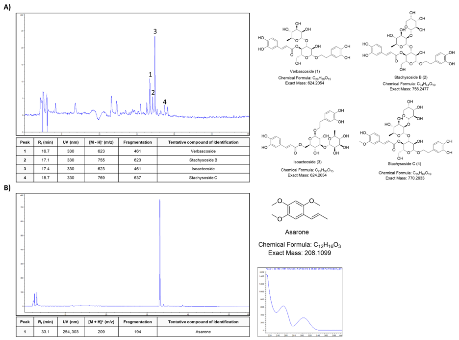
We used HPLC and LC-MS/MS with ESI to characterize the AGS and SSM extracts. Each major peaks were identified in the HPLC profile of the AGS and SSM extracts. The identification of the chemical compounds was also carried out by comparing the molecular ion peaks along with the MS fragmentation pattern with those of the literature [20]. As shown in (Figure 1A), AGS extract Peaks 1, 2, 3, and 4 were tentatively identified as Verbascoside, Stachysoside B, Isoacteoside, and Stachysoside C, respectively. In addition, SSM extract major peak was tentatively identified as Asarone (Figure 1B).

biomedres-openaccess-journal-bjstr

Figure 1: Fingerprint analysis of AGS and SSM extracts. HPLC and LC-MS/MS analysis of the major compounds from

(A) AGS and

(B) SSM extracts, respectively.

Mortality Rate and LC50 Value

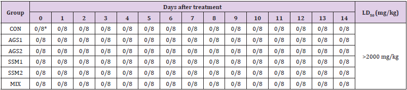
The results of toxicity signs and mortalities caused by SSM, AGS, and MIX in ICR mice are presented in (Table 1). From the result of the 14-day observation period of the treatment group receiving a single oral administration of 1,000 and 2,000 mg/kg doses of AGS, SSM, and MIX and the control group receiving a single oral administration of sterile physiological saline, no mortalities were noted in all groups, including the highest dose group. Therefore, the minimum lethal dose of AGS, SSM, and MIX exceeds 2,000 mg/kg in ICR mice. In addition, lethal concentration 50 (LC50) of AGS, SSM, and MIX is estimated to be over 2,000 mg/kg.

biomedres-openaccess-journal-bjstr

Table 1: Mortality of ICR mice orally administered with AGS, SSM, and MIX.

Note: CON; Control group, AGS1; AGS 2,000 mg/kg (day) medication group, AGS2; AGS 1,000 mg/kg (day) medication group, SSM1; SSM 2,000 mg/kg (day) medication group, SSM2; SSM 1,000 mg/kg medication group, MIX; MIX 2,000 mg/kg (day) medication group. *Values are expressed as Number of dead animals/ Number of animals examined.

Drinking, Feed Intake and Clinical Signs

After comparing the changes in drinking yield and feed intake by single oral administration of AGS, SSM, and MIX in the treatment groups with the control group receiving a single oral administration of sterile physiological saline, administration of the test substances did not result in any significant differences in the changes in drinking yield and feed intake (data not shown). Further, no abnormal findings in clinical symptoms related to single oral administration of sterile physiological saline, AGS, SSM, and MIX were observed, including hair loss, activity decline, gait disorder, behavior disorder, squat, diarrhea, swelling, dyspnea, grooming, jumping, tearing, lethargy, polyuria, vomiting, nasal discharge, numbness, suppleness, etc. (data not shown).

Changes in Bodyweight

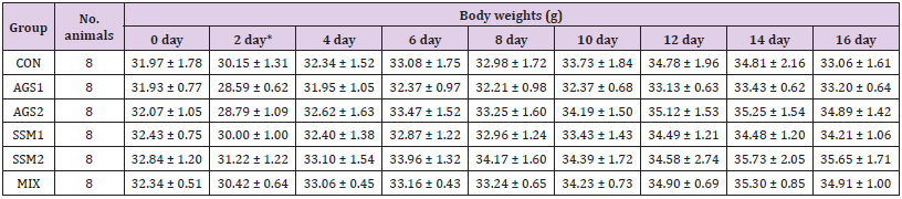
The results of changes in bodyweight of the treatment and control groups are presented in (Table 2). After oral administration, normal weight gain over time was observed in the AGS, SSM, and MIX administration groups and the control group compared to the weight before administration. No significant weight change was noted after administration, compared to that before administration, in the treatment (AGS, SSM, and MIX administration) and control groups, indicating no toxicity.

biomedres-openaccess-journal-bjstr

Table 2: Body weights changes of ICR mice orally administered with AGS, SSM, and MIXb.

Note: CON; Control group, AGS1; AGS 2,000 mg/kg (day) medication group, AGS2; AGS 1,000 mg/kg (day) medication group, SSM1; SSM 2,000 mg/kg (day) medication group, SSM2; SSM 1,000 mg/kg medication group, MIX; MIX 2,000 mg/kg (day) medication group. The data are presented as mean ± standard deviation. *Day after AGS, SSM, and MIX administration.

Necropsy Results and Change in Organ Weight

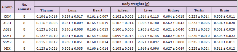
The results of gross findings on major organs by necropsy of all ICR mice after the 14-day observation period are presented in (Table 3). There were no gross abnormalities or abnormal lesions on major internal organs suspected of causing abnormalities by the administration of test substances in all animals in the control and treatment groups. In addition, no significant changes in the weights of the thymus, lungs, heart, spleen, liver, kidney, testes, and brain were observed in the treatment groups compared with that in the control group.

biomedres-openaccess-journal-bjstr

Table 3: Organ weights of ICR mice orally administered with AGS, SSM, and MIX.

Note: CON; Control group, AGS1; AGS 2,000 mg/kg (day) medication group, AGS2; AGS 1,000 mg/kg (day) medication group, SSM1; SSM 2,000 mg/kg (day) medication group, SSM2; SSM 1,000 mg/kg medication group, MIX; MIX 2,000 mg/kg (day) medication group. The data are presented as mean ± standard deviation.

Hematological Analysis

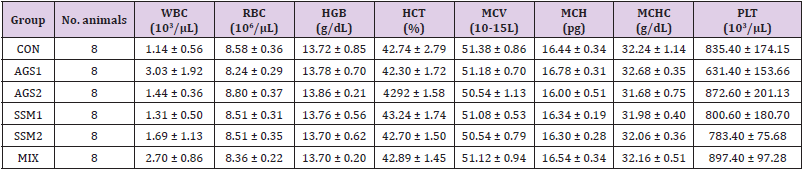
The evaluation of WBC, RBC, HGB, HCT, MCV, MCH, MCHC, and PLT using a hematological analyzer was used to investigate hematological changes 14 days after oral administration of either sterile physiological saline, AGS, SSM, or MIX. The results of this evaluation are shown in (Table 4). From the results of hematological analysis on collected whole blood from the treatment and control groups, PLT in the groups treated with AGS, SSM, and MIX showed slight reduction compared to the control group, albeit not significantly. The other categories showed no significant changes between the control group and the treatment groups.

biomedres-openaccess-journal-bjstr

Table 4: Hematological analysis of ICR mice orally administered with AGS, SSM, and MIX.

Note: CON; Control group, AGS1; AGS 2,000 mg/kg (day) medication group, AGS2; AGS 1,000 mg/kg (day) medication group, SSM1; SSM 2,000 mg/kg (day) medication group, SSM2; SSM 1,000 mg/kg medication group, MIX; MIX 2,000 mg/kg (day) medication group. The data are presented as mean ± standard deviation.

Blood Chemistry Analysis

The results of serum ALT, AST, BUN, CREA, and LDH values measured using an automated blood chemistry analyzer for investigating blood biochemical changes after 14 days in the treatment and control groups are shown in (Table 5). It was found that a single oral administration of AGS, SSM, and MIX induced a slight change in the test parameters, but in general, no significant changes were observed in all indicators between the control group and the treatment groups.

biomedres-openaccess-journal-bjstr

Table 5: Blood chemistry analysis of ICR mice orally administered with AGS, SSM, and MIX.

Note: CON; Control group, AGS1; AGS 2,000 mg/kg (day) medication group, AGS2; AGS 1,000 mg/kg (day) medication group, SSM1; SSM 2,000 mg/kg (day) medication group, SSM2; SSM 1,000 mg/kg medication group, MIX; MIX 2,000 mg/kg (day) medication group. The data are presented as mean ± standard deviation.

Discussion

Recently, various types of medicines are being used, but problems such as adverse effects due to toxicity also appear. Not only is the interest in functional foods and natural medicines using herbal medicines and crude drugs is increasing worldwide but also their effect and efficacy are being verified, owing to an increasing demand for various forms of natural-product derived pharmaceuticals [21,22]. However, in the general practice of natural medicine, which prescribes a combination of various crude drugs, exact ingredients and specifications are not well established and data on their safety and toxicity are often insufficient, thereby necessitating specific and accurate information on them [6,7,23]. Therefore, in this study, to obtain an objective basis for the safety of AGS, SSM, and MIX and experimentally evaluate their acute toxicity, the observation of clinical symptoms, necropsy findings, mortality, and weight change, and hematological analysis were conducted after administering the test substance to ICR mice. First, an acute toxicity test was performed to confirm the safety of AGS, SSM, and MIX, following which all subjects in the treatment groups treated with AGS, SSM, and MIX as well the control group treated with sterile physiological saline showed no mortality and no significant change in body weight.

Therefore, based on the US EPA standards that classify a substance safe if its LD50 value by oral administration is >2,000 mg/ kg, AGS, SSM, and MIX are considered to be very safe in terms of acute toxicity. Next, from the result of gross examination and organ weight measurement by necropsy to confirm the effects of AGS, SSM, and MIX on the major internal organs, no gross abnormalities or abnormal lesions were observed and no significant changes in the weights of major organs, such as the thymus, lungs, heart, spleen, liver, kidney, testes, and brain, were observed. In general, in a single-dose toxicity study, if gross abnormalities in organs or tissues are observed, histopathological examination should be performed. However, in this study, histopathological examination was not performed as no gross abnormalities were observed in all experimental animals. Also, hematological and blood chemistry analyses using whole blood and serum, respectively, collected at the end of the observation period revealed slight changes in some test parameters, but in general, no significant changes were noted in terms of AGS, SSM, and MIX treatment in all test parameters. In summary, as AGS, SSM, and MIX did not show any acute toxicity on the test animals, they could be considered relatively safe for oral administration. In addition, they can be expected to be used as natural materials without acute toxicity through further investigation of their physiological effects.

However, there are some limitations of determining the toxicity of natural herbal medicines through only a single oral administration acute toxicity study. Hence, it is necessary to conduct repeated oral administration toxicity studies for additional 2 or 4 weeks and 13 weeks (long term) and genotoxicity studies subsequently. In addition, further research on human safety evaluation is essential, based on which more precise and scientifically accurate safety data can be obtained by establishing systematic toxicity information on AGS, SSM, and MIX.


For more Articles on: https://biomedres01.blogspot.com/
Antimalarial Aloe Compounds

  Antimalarial Aloe Compounds Introduction Among the most prevalent diseases caused by protozoan parasites, malaria is caused by parasites o...