Thursday, April 25, 2024

Discrepancies of Pediatric Data of BNT162b2 mRNA Covid-19 Vaccine in the Assessment Report of the European Medicines Agency


Discrepancies of Pediatric Data of BNT162b2 mRNA Covid-19 Vaccine in the Assessment Report of the European Medicines Agency

Clinical Efficacy

The Assessment Report of pediatric study of BNT162b2 mRNA Covid-19 Vaccine (Comirnaty) says that there was no cases of Covid-19 in 1119 adolescents 12 to 15 years of age administered vaccine (0%) and there were 18 cases in 1110 (1.6%) of those administered placebo [1]. The vaccine efficacy (Relative Risk Reduction, ARR) is 100% while the Absolute Risk Reduction (ARR) is 1.6 % (18/1119 – 0/1110) [1]. Literally, BNT162b2 mRNA Covid-19 Vaccine prevented mild Covid-19 in 1.6% of the study population. At the cut-off date, no severe cases and no deaths were reported in 12 15 years-old age group1, i.e., no benefit in respect to prevention of severe or death Covid-19 was demonstrated. Prevention of long-term Covid-19 was not investigated.

Safety: Serious Adverse Events

The Assessment Report says that the rate of Serious Adverse Events (SAEs) in adolescents in the time frame from dose 1 to one month after dose 2 (until the data cut-off, 13 March 2021) was very low and similar between vaccine and placebo arms (≤0.4%).1 However, it is a misleading statement:

1) Mixing an active drug arm and a control arm is unacceptable from a scientific point of view. Indeed, there were 5 serious adverse events (SAEs) in the vaccine arm and 2 SAEs in the placebo arm; serious psychiatric events were reported in 4 subjects in the vaccine arm and 0 in the placebo arm [1].

2) In addition, 2 adolescents originally randomized to the placebo had life-threatening SAEs after they turned 16 years during the study and were unblinded to receive BNT162b2, [1]

3) Frequency category “very low” is not consistent with the Medical Dictionary for Regulatory Activities (MedDRA). According to MedDRA, the SAEs should be classified as uncommon (frequency ≥1/1,000 to <1/100).

5 above-mentioned SAEs in adolescents of the BNT162b2 group included [1]:

1) Participant No 1 had neuralgia with 3 emergency room visits beginning day 1 after dose 2 and subsequently, abdominal pain and constipation. She was diagnosed with functional abdominal pain, referred to psychology and physical therapy after which symptoms were reported as gradually improving.

However, SAEs persisted after 1-month post Dose 2 and remained unresolved at the data cut-off (see follow up below),

2) Participants No 2 and 3 had depression,

3) Participant No 4 had concurrent anxiety and depression,

4) Participant No 5 had concurrent appendicitis and focal peritonitis.

2 adolescents originally randomized to the placebo group had life-threatening SAEs after they turned 16 years of age and were unblinded to receive BNT162b2 [1]:

1) Participant No 6: an anaphylactoid reaction reported 3 days after receiving the first dose of BNT162b2 with a duration of 1 day, leading to study withdrawal,

2) Participant No 7: a depression reported 7 days after receiving the first dose of BNT162b2 reported as ongoing/resolved at the time of data cut-off date.

SAEs reported from after 1-month post Dose 2 up to the data cutoff date included suicidal ideation and appendicitis (each appearing in 1 participant) [1]. In participants administered placebo 2 SAEs have been reported [1]. Follow-up data, consistent with Participant No 1, was presented on 28 June 2021 during a meeting organized by Senior United States Senator from Wisconsin Ronald Harold Johnson. Stephanie de Garay described the experiences of her daughter Maddie de Garay after the 2nd dose of the Pfizer Covid-19 vaccine in the pediatric study. Upon receiving her second dose on January 20th, over the next 24 hours, Maddie developed severe abdominal and chest pain. She had extreme pain in her fingers and toes making them turn white and cold. Later, abdominal, muscle, and nerve pain became unbearable. Additional symptoms included gastroparesis, nausea and vomiting, erratic blood pressure and heart rate, memory loss. She mixed up words, had brain fog, headaches, dizziness, fainting, and then seizures, developed verbal and motor tics. She lost feeling from the waist down and got muscle weakness, drastic changes in her vision, urinary retention and loss of bladder control, severe irregular menstrual cycles, and eventually got a nasogastric tube for her nutrition [2].

Discussion

By article 14-a (1) of the Regulation (EC) 726/2004, conditional marketing authorization for medicinal products intended for the treatment or prevention of seriously debilitating or life-threatening diseases may be granted before the submission of comprehensive clinical data [3]. However, Covid-19 is neither a debilitating nor life-threatening disease in the majority of adolescents [4]. Severe cases occur rarely, and predominantly in subjects with underlying conditions, adolescents with risk of severe disease due to underlying conditions were not specifically studied [1]. The study did not demonstrate any benefit in respect to the prevention of severe Covid-19. It demonstrated more SAEs instead: 5 adolescents in the BNT162b2 group reported any SAE from Dose 1 to the data cut-off date up to 1 month after Dose 2. Two adolescents originally randomized to the placebo group had life-threatening SAEs after they turned 16 years during the study and were unblinded to receive BNT162b2. These SAEs are not included in the benefitrisk assessment of the Assessment Report [1]. By the report of the Centers for Disease Control and Prevention from 1 March 2020 to 24 April 2021, the cumulative Covid-19–associated adolescent hospitalization rate was 49.9 per 100,000.2 If we assume that vaccines prevent all cases of severe Covid-19 (49.9/100,000), the absolute risk reduction is 0.05%, i.e., 2000 vaccinations might prevent 1 hospitalization. This means that effectiveness in respect to the risk of hospitalization was about 10 times lower than the frequency of SAEs (by definition, SAE results in death, is lifethreatening, requires hospitalization or prolongation of existing hospitalization, results in persistent or significant disability or incapacity, or a congenital anomaly/birth defect) [4].

COVID-19 pandemics did not increase pediatric mortality in EUROMOMO countries. Instead, pediatric mortality steadily decreased from spring 2020 until June 2021, i.e., beginning of the pediatric vaccination against COVID-19 [5]. Time relation does not mean causation but still should be considered. The pediatric study did not provide data to what extent vaccination provides protection against asymptomatic infection, and whether vaccination prevents further transmission.1 The pediatric data came from the period before the emergence of the Delta variant. There are no bridging studies to extrapolate the pediatric data to today’s situation. The power of the pediatric study was neither sufficient to demonstrate any benefit in terms of reduction of the risk of severe COVID-19 or death nor the risk of serious or life-threatening adverse reactions. It should also be noted that the BNT162b2 mRNA Covid-19 vaccine has been used for mass vaccination in children and therefore any doubt regarding safety or efficacy is unacceptable [6,7].

Conclusion

1) Covid-19 should not be considered a serious, debilitating, or life-threatening disease in 12 to 15-years-old adolescents, i.e., conditions of the conditional marketing authorization are not met.

2) Known benefits of the BNT162b2 vaccine in 12 to 15-years-old adolescents do not exceed known risks. In addition, long-term risks are unknown.


For more Articles on: https://biomedres01.blogspot.com/

Wednesday, April 24, 2024

My Therapy Concept from 25 Years of Experience in Dental Sleep Medicine


My Therapy Concept from 25 Years of Experience in Dental Sleep Medicine

Introduction

My therapy concept is based on the idea of using three highly precise aids, that exhibit very little redundancies. The combination of these aids yields a significant increase in quality, effectiveness, and efficiency of the entire treatment process. The aids I am referencing here can be split into three separate solutions: first the vector diagram as a framework, secondly the JS-Gauge® as an instrument and thirdly the F-UPS® as MAD (mandibular advancement device). In our clinic, the combination of these aids has significantly reduced laboratory and treatment times and increased our patients’ satisfaction.

Risk Profiling (Vector Diagram)

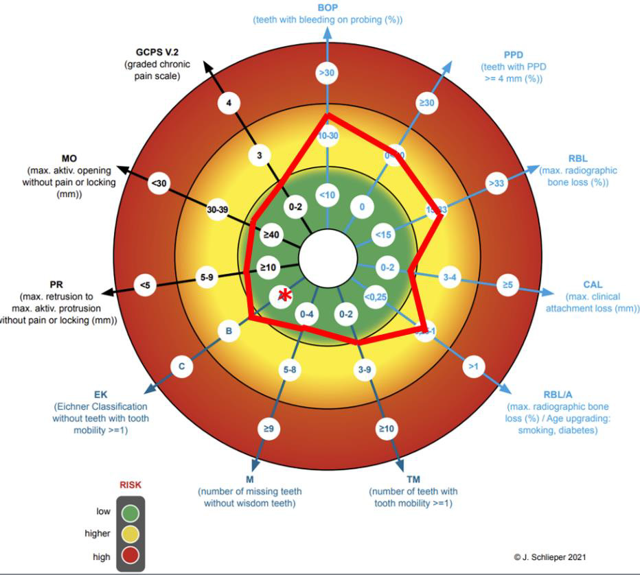
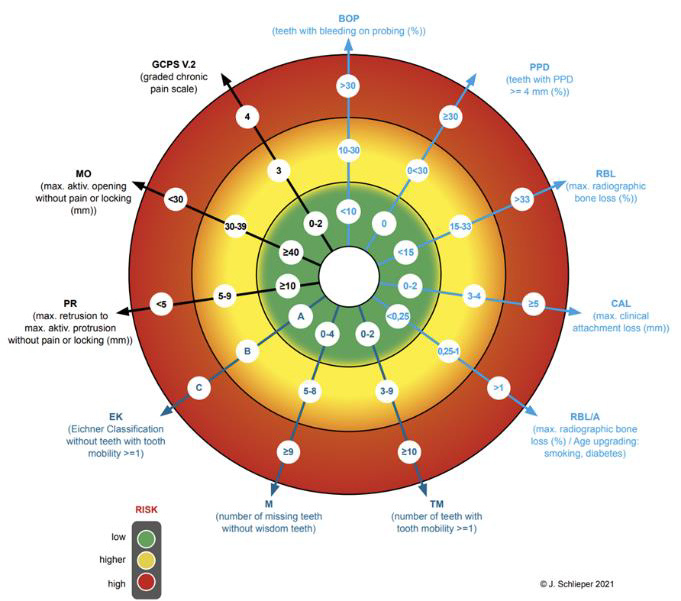
Both at the beginning and throughout the MAD therapy [1,2] it is critical to measure specific indicators to help guide decisionmaking. Thus, we can effectively weigh risks and opportunities of the MAD therapy against one another and craft an effective treatment plan. A comprehensive and tailored risk profiling [3] is a viable solution here and creating a framework to standardize the process of identifying these so-called predictors can greatly increase efficiency [4]. Said framework which i developed can be seen in Figure 1 in the form of a vector diagram, which allows us to visually compose a risk profile for each patient. This visual representation further allows for a swift comparison between consecutive risk assessments, to allow for the evaluation of progress throughout the treatment [5]. The predictor values are segmented into three risk groups and color-coded via a traffic light system (red: high risk; yellow: moderate risk; green: low risk).

biomedres-openaccess-journal-bjstr

Figure 1.

biomedres-openaccess-journal-bjstr

Figure 2.

Utilizing this visual aid allows for a quicker assessment that can be intuitively grasped by both the practitioner and the patient. This not only facilitates the interdisciplinary decision-making between the dentist and sleep specialist, but also aids the dentist with educating the patient, thus also informing the patient’s decisionmaking process. Moreover, the vector diagram helps guide the therapeutic decision-making throughout the entire MAD therapy. Therefore, the framework helps to both optimize the starting conditions for the MAD therapy and reduces the risks throughout the therapy. However, this diagram is not intended for the precise recording of all dental parameters. Nor shall it be used to assess the quality of oral health overall. The diagram’s intention is to primarily increase the quality, effectiveness, and efficiency of the therapeutic decision-making process for MAD. Above all, it aims to lay the foundation for treating large patient populations and thus making it a viable tool for the use with public health insurance providers. The risk assessment covered by the vector diagram spans the three areas of biological, mechanical, and functional values of the stomatognathic system.

The biological area relates to periodontics [6], the mechanical area to prosthetics [7] and the functional one to the function [8]. The vectors and the labelling of these 3 areas are shown with different colors light blue, dark blue and black for better visual differentiation. The collected values are marked in the diagram, either with its value in the given selection or scaled on the vector ray. This makes it easier to identify minor changes during a followup. The line connection results in the individual biological risk profile for the MAD therapy. The mechanically relevant findings for tooth loosening, the number of teeth and the profile of the support according to the Eichner classification are also collected. The Eichner classification is subdivided into the profile of the dental or implant logical support and the profile of the occlusal support. The Eichner classification is basically only a classification of the gap dentition according to the profile of the dental support zones intended for prosthetic therapy. However, this classification is not sufficient here for the risk analysis in MAD therapy. In the vector diagram, this classification is therefore further subdivided into a dental and implantological support zone profile without prosthetic gap closure, as well as the occlusal support zone profile at the time of the examination, possibly with prosthetic gap closure. The dental classification is marked without, while the occlusal classification is marked with an asterisk in the vector diagram (Figure 2).

Finally, the 3 relevant functional findings, which include active degree of protrusion, active degree of mouth opening and the graded chronic facial pain after DC/TMD need to be assessed. The active degree of protrusion is measured from the maximum retrusion into the maximum protrusion in the lying position after three attempts. In my opinion and in the opinion of many other colleagues, this is the most precise measurement method. Only values that can be achieved without locking of the jaw or pain are measured. The same procedure is used to measure the active mouth opening. Finally, the GCPS grade is entered from the assessment of the corresponding questionnaire according to DC/TMD, which completes the creation of the risk profile for the function. In (Figure 2) you can see an example of a fully completed primary assessment.

Adjustment and registration with bite gauges (JS-Gauge®)

Once the decision for or against MAD therapy and for or against preparatory measures has been made and these have been completed, the second, most important treatment step follows: the adjustment and registration of the starting position, which is critical to produce a MAD [2].

From January 1, 2022, onwards the MAD therapy will be covered by public health insurance in Germany and the requirements to receive the needed coverage include an individual three-dimensional registration of the starting position [9]. This registration must be tailored to the individual circumstances of the patient as well as to the individual design of the MAD that is to be used. Bite gauges are used as aids for this [10,11]. They offer the advantage of flexible adjustments under functional, neuromuscular conditions when compared to the use of no bite gauge or the axiographs. All bite gauges currently on the market neither fully nor ideally meet these requirements. Bite gauges, which are held between the rows of teeth by the patient biting down, are subject to variability during adjustment and registration, which reduces precision. The precision can be increased considerably by securely fixing the bite gauge to the upper jaw [12]. A bite spoon is fixed to the upper jaw with A-silicone and the lower jaw is supported by means of a support pin registry.

biomedres-openaccess-journal-bjstr

Figure 3.

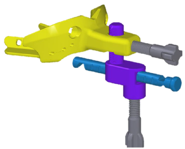
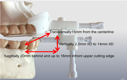
This allows for more comfort due to a relaxed jaw, interferencefree adjustments and a registration in the lying treatment position without a forced bite. The JS-Gauge® (Figure 3) which i developed consists of the bite spoon, in yellow, the vertical pin, here in turquoise, and the horizontal pin, marked in blue, as well as two fixing screws, colored gray. A standout feature of the JS-Gauge® is that it can be continuously adjusted in all three spatial dimensions sagittally, vertically and transversely. The support of the horizontal pin on the tooth edges of the mandibular incisors, can be continuously adapted for a safe adjustment of the mandible on the JS-Gauge®. The adjustment and registration range (Figure 4) extends sagittally from 20 mm behind and up to 16 mm in front of the upper edge of the incisor. A vertical bite lock can be set from 2,0 mm IID to approx. 14 mm IID. Transversally, the JS-Gauge® allows adjustment 15 mm from the centerline. This transversal adjustment should also be adjusted habitually in a lying position without functional symptoms occurring. We want the patient to be as relaxed as possible.

biomedres-openaccess-journal-bjstr

Figure 4.

biomedres-openaccess-journal-bjstr

Figure 5.

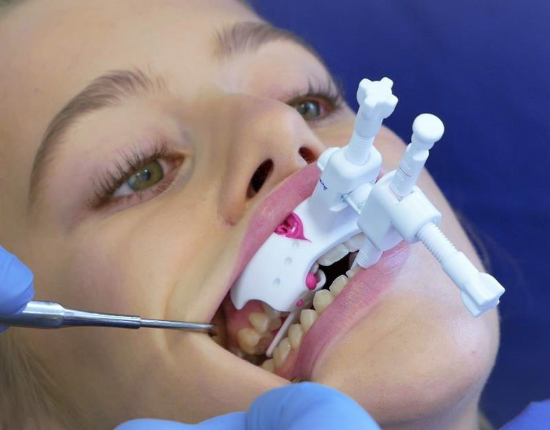
The adjustment with the bite gauge should be tailored to the individual patient’s situation and the design of the MAD [13,14]. With increasing bite blockage with the same protrusion, which can be measured as inter incisor distance (IID) in relation to the upper jaw, the load on the craniomandibular system increases [15]. We can gather from this that the percentage of the same protrusion in relation to the maximum protrusion increases with increasing bite blockage [16]. In this respect, registration with bite gauges should be carried out with as little bite blockage as the bite conditions, the functional findings and the MAD Design allows. The protrusion should be adjusted from the maximum retrusion to approx. 5 mm in the comfort zone of the patient. I recommend a 5 - 10 minute control of this position with regard to the functional findings and the comfort findings of the patient. The 3D adjustment of the starting position is complete if no pathological functional findings occur, and the patient does not complain about discomfort within 5-10 minutes (Figure 5). After use, the JS-Gauge® is reprocessed with the help of the tray and the box. The reprocessing meets the requirements of the MDR (Medical Device Regulation, EU 2017/745) in all stages.

MAD (F-UPS®)

From this starting position, sleep medical titration continues after the MAD has been inserted [2,9]. In order to be able to achieve the lowest possible bite blockage, especially in patients with a deep bite or a flat Spee curve, I developed a MAD (H-UPS®) as early as 1997 [17] that enables this. This leaves the front teeth mostly free. This makes the plastic construction more unstable, yet this instability is absorbed by a steel arch to which the Herbst hinges are attached by laser welding. Although this construction has many advantages, the disadvantage is the mechanics of the Herbst hinge. As every time the patient opens his mouth during the night, a retrusion occurs, frontal elastic bands are intended to prevent this but unfortunately cannot do so completely. If the Herbst telescope is mounted on the MAD in reverse, unphysiological stresses arise on both the MAD and the dental arches as well as on the craniomandibular system when the mouth is opened. Therefore, no desired physiological conditions can be achieved. Inclined planes attached to the side also do not allow a physiological opening of the mouth and do not reliably prevent an opening of the mouth. Frontal elastics are also required here. As we can see all these approaches are at least somewhat flawed.

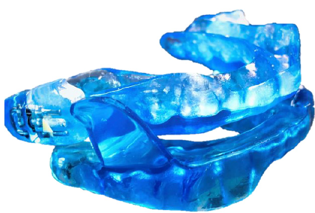
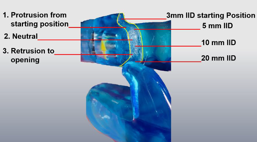
In my opinion, the solution lies in the fin-like design with the lateral guide elements. I developed a MAD (F-UPS®) in 2018 (Figure 6) that enables this. With these it is possible for the typically mouth openings up to 10 mm IID [18] to physiologically occur during sleep without retrusion, which constitutes a physical adjustment of the guide way of the lateral elements without any loss of effectiveness of the MAD. The guide way of the lateral elements is divided into three parts (Figure 7). From the starting position, the first part starts with a jaw opening with a protrusion movement of up to approx. 5 mm. The protrusion is intended to reflexively prevent the number of further jaw openings with a larger IID during sleep. If there is still another jaw opening, the second part of the movement follows with a jaw opening with unchanged protrusion up to 10 mm IID. Only from 10 mm IID, which normally does not occur during sleep [18], is 3. the further jaw opening with retrusion up to the disconnection of the two splint parts with release for maximum jaw opening. The fins of the F-UPS® are not made separately. In this design, the upper and lower jaw splints are CAD designed and milled from a single block. This is the only way to produce the F-UPS® with this fin design for an optimized function without retrusion and with the ideal recess in the anterior tooth area for the least possible bite blockage with very high stability.

biomedres-openaccess-journal-bjstr

Figure 6.

biomedres-openaccess-journal-bjstr

Figure 7.

Optionally, this sagittal guideway of the lateral elements can be made more precise from the starting position by a further registration in 10 mm IID to be able to shape the guideways transversely. This is e. g. recommended if the jaw opening-path shows a lateral deviation. For this purpose, after the first registration of the starting position, the sagittal, vertical, and transversal setting values are adopted from the JS-Gauge® the IID setting is increased to 10 mm and readjusted, if necessary, according to functional aspects. After frontal and lateral coating with A-silicone, the JSGauge ® is removed. This procedure is possible because the jaw relation can be transferred from a first registration to the second registration by adopting the 3D setting values of the JS-Gauge®. It is therefore not necessary to keep registrations for later new MAD productions. Knowledge of the 3D setting values of the JS gauge is sufficient for this.

Conclusion

During the development of these three aids, the treatment of my patients has steadily improved and due to the use of these aids in conjunction with each other the treatment quality has improved yet again.


For more Articles on: https://biomedres01.blogspot.com/

Monday, April 22, 2024

Healing and Prophylactic Efficacies of Topical Composite Herbal Medication on Overuse-induced Achilles Tendinosis


Healing and Prophylactic Efficacies of Topical Composite Herbal Medication on Overuse-induced Achilles Tendinosis

Introduction

Tendinopathy is a clinical condition characterized by pain and swelling in and around degenerative tendons [1]. Achilles tendon is vulnerable to developing tendinopathy, especially among athletes due to the continuous, prolonged and intense functional demands imposed on it during sports training and competitions [2]. Tendinopathy affects not only athletes but 25% to 30% of injured were non-athletes resulting in significant loss of working days [3]. Hence, this condition poses a heavy toll on the individual as well as the society by increasing workers’ compensation costs. Traditionally, Achilles tendinopathy was described as Achilles tendinitis which refers to an inflammatory process with the occurrence of pain and swelling at the tendon. With more scientific evidence gathered, there is better understanding to the condition that it is a degenerative process without histological or clinical signs of intratendinous inflammation [4,5], the condition is now described as Achilles tendinosis which is the outcome of imbalance between degeneration and synthesis of cell-matrix upon repetitive trauma [6]. However, the etiology behind Achilles tendinosis is still unclear but various factors have been reported to induce the condition which included overuse, decreased blood supply and muscle imbalance or weakness [7]. Essentially, Aström [8] reported a survey of surgical and histopathologic findings that 90% of biopsy specimens from symptomatic parts of tendons with tendinopathy exhibited symptoms of tendinosis including abnormal fiber structure, focal hypercellularity and vascular proliferation [8]. This suggested chronic degeneration of Achilles tendon would be a risk factor for rupture of the tendon. Hence, in addition to treatment upon occurrence of injuries, prophylaxis against development of tendinosis is important for reducing risk of tendon rupture as well.

Currently, majority of the studies on management of Achilles tendinosis focused on the therapeutic effects of the treatment protocols upon occurrence of the disorder. For instance, Furia [9] demonstrated that high-energy extracorporeal shock wave therapy (ESWT) was more effective in treating chronic insertional Achilles tendinopathy in terms of improved perceived condition of diseased Achilles tendon and pain score [9]. However, in a double-blinded randomized controlled trial by Costa et al, no significant treatment effect was found in 49 patients with Achilles tendinosis treated with low energy ESWT [7]. Nevertheless, a recent placebo-controlled randomized control trial which compared the application of pointfocused and line-focused applicator in the treatment of achilles tendinopathy, found significant improvement for all study groups (including placebo group) without a statistically significant benefit for ESWT groups during 24 weeks [10]. Besides, Stergioulas, et al. [11] reported that low-level laser therapy (LLLT) with eccentric exercise would decrease pain, morning stiffness, tenderness to palpation, and improve active dorsiflexion and crepitus with no side effects in patients with Achilles tendinosis as compared to those underwent eccentric exercise only [11]. In addition to physical treatments, healing effects of medications have also been studied. Paoloni, et al. [12] demonstrated that topical glyceryltrinitrate (GTN) significantly reduced pain, tenderness on palpation and improved ankle plantar flexor activities in subjects with non insertional Achilles tendinopathy [12].

Complementary to the western treatment approach, Traditional Chinese herbal medicine (TCM) has long been used for treating tendon and ligament injuries. Fu, et al. [13] demonstrated the ultimate strength of healing tendon was significantly promoted and collagen deposition was enhanced with better fiber alignment being observed in rat patella tendons after injection of total flavones of Hippophae rhamnoides, also known as Shaji [13]. Further, our previous findings suggested that topical application of Panax notoginseng extract coupled with therapeutic ultrasound would improve the strength of repairing rat ligament [14]. Although previous works had demonstrated the therapeutic potential of TCM on tendon repair, information on treatment effect as well as the prophylactic potential of TCM on overuse-induced tendinosis is scarce. Thus, the objective of the current study was to investigate the healing effects and prophylactic efficacy of a composite TCM extract on overuse-induced Achilles tendinosis.

Materials and Methods

Chinese Herbal Medication

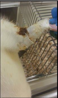
The herbal application formula for this study comprised Dipsaci Radix (DR), Rhizoma Notoginseng (RN), Flos Carthami (FC), and Rhizoma Rhei (RR) in the ratio 1:1:1:1. These herbs were reported to control inflammation, stimulate circulation and promote fibroblastic activity for tissue repair [14-17]. After purchasing the raw herbs, their identities were authenticated using thin-layer chromatography with reference to the methods recommended by the Chinese Pharmacopoeia (2010). The herbs were crushed into small pieces and extracted with distilled water twice by heating under reflux followed by heating under reflux with 95% ethanol twice. The collected aqueous and ethanolic extracts were filtered and lyophilized to give the powder. The extraction yields of aqueous and ethanolic extracts were 40.0% and 5.0% respectively. The herbal paste was prepared by mixing 3.9g of aqueous extract and 0.5g of ethanolic extract in 3.4ml 50% ethanol-water. The herbal paste was applied topically at the location of Achilles tendon of both hind limbs and wrapped with Micropore surgical tape (3M, USA) (Figure 1). The herbal plasters were replaced on daily basis.

biomedres-openaccess-journal-bjstr

Figure 1: Topical application of herbal paste at rat Achilles tendon.

Animal Model

Forty 3-month-old female Sprague-Dawley (SD) rats were used in this study. The animals were randomly assigned into 4 groups, namely, Cage control (CC) which received no treatment, Exercise without herbal treatment (EX), Exercise with herbal treatment provided before forced running period (Pre-HB) and Exercise with herbal treatment provided after forced running period (Post-HB), with 10 rats in each group. Overuse-induced Achilles tendinosis was created in EX Pre-HB and Post-HB groups by an enforced running protocol [18]. Briefly, the rats were subject to run on a rat treadmill (Duen Production BCPT-98, Hang Zhou, Zhejiang, China) at 20m/ min with a 20-degree decline slope modified from the eccentric loading model of Soslowsky [19]. In order to increase loading on the rats’ hind limbs, the upper trunk of rats was suspended with a harness so that their forelimbs were lifted off and hence the rat ran in a bipedal pattern with the hind limbs. The rats were subject to the running exercise for 1 hour per day, 7 days per week for 8 weeks. During the 4 weeks before forced exercise period, Pre-HB rats were treated with herbal extract application to both Achilles tendons on daily basis whereas CC and EX rats were kept in cage without any treatment. Upon completion of the 8-week running program, the Post-HB rats were treated with herbal extract application to both Achilles tendons on daily basis for 6 weeks. Pre-HB, EX and CC animals were sacrificed by CO2 inhalation at the end of forced exercise period whereas Post-HB rats were sacrificed at the end of the 6-week herbal treatment period. Both Achilles tendons were harvested and kept frozen at -70°C until further testing. The protocol of the current study was approved by the Animal Ethics Review Committee of the administering institution.

Histological Analysis

The left Achilles tendons were used for histological analysis. The specimens were rehydrated in 0.01M phosphate-buffered saline (PBS) containing 20% sucrose for 30 minutes at room temperature. Then, the tendon samples were sectioned at a thickness of 7μm with a cryostat followed by hematoxylin-eosin (H&E) staining. The sections were examined using light microscope (Model: Eclipse 80i, Nikon, Japan) immediately after staining. Images of the sections were digitally captured with a camera (Model: SPOT Flex 15.2, 64Mp, SPOT Imaging Solutions, Sterling Heights, MI, USA) mounted on the microscope at 100x magnification. The captured images were further analyzed using ImageJ version 1.46r (NIH, USA). The level of cellularity was estimated based on the density of cell count in the area of tendon captured.

Biomechanical Testing

The left Achilles tendons were used for histological analysis. The specimens were rehydrated in 0.01M phosphate-buffered saline (PBS) containing 20% sucrose for 30 minutes at room temperature. Then, the tendon samples were sectioned at a thickness of 7μm with a cryostat followed by hematoxylin-eosin (H&E) staining. The sections were examined using light microscope (Model: Eclipse 80i, Nikon, Japan) immediately after staining. Images of the sections were digitally captured with a camera (Model: SPOT Flex 15.2, 64Mp, SPOT Imaging Solutions, Sterling Heights, MI, USA) mounted on the microscope at 100x magnification. The captured images were further analyzed using ImageJ version 1.46r (NIH, USA). The level of cellularity was estimated based on the density of cell count in the area of tendon captured.

Biomechanical Testing

The right Achilles tendons were tested for their biomechanical properties. Each specimen was dissected by leaving only the intramuscular tendinous fibers, Achilles tendon and calcaneus intact. The intramuscular tendinous fibers were then secured between 2 plastic strips with epoxy glue (Aron Alpha, Toagosei Co. Ltd, Columbus, OH) and mounted onto the cross-heads of a material testing machine (Model: Synergie 200, MTS System Corporation, Ivry sur Seine Cedex, France). An extensometer (Model: 634.12F- 24, MTS System Corporation, Eden Prairie, MN) was attached to the margin of the cross-heads for measuring the local strain of the tendon. The specimens were kept moist with normal saline during the entire testing process. The specimen was pre-conditioned with 10 oscillation cycles of 2.5% strain at 10mm/min to minimize the effect of deep freezing [20], and then stretched to 2.5% strain and maintained for 5 minutes. The load required to sustain the length of specimen was recorded at 5 Hz throughout the test. The percentage change in load reflected the load-relaxation property. The specimen was then subject to ultimate tensile failure testing at a strain rate of 500mm/min with data sampling rate of 50Hz [14]. The ultimate failure load (UFL) was defined as the maximum load before failure and the structural stiffness was defined as the gradient of the linear portion of the load-deformation curve.

Statistical Analysis

Statistical analysis was performed using SPSS ver.20 (IBM, USA). Results were presented as means and standard deviations. Intergroup differences in biomechanical results and cell densities were evaluated with one-way ANOVA with post-hoc LSD test. Statistical significance was set at p<0.05.

Results

Histological Analysis

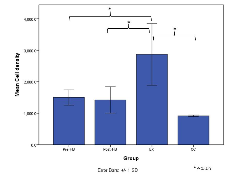
After 8 weeks of bipedal downhill running exercise program, when compared to CC group (Figure 2a), hypercellularity was observed in all tendon specimens of EX group through intra-group variation at the level of cellularity existed (Figure 2d). Besides, mild level of hypercellularity was found in some of the tendon specimens of Pre-HB and Post-HB groups (Figures 2b & 2c). The average density of cell count of EX group (2868.7±979.3 /mm2) was significantly higher than those of CC group (918.2±24.4 /mm2) (p=0.002, post-hoc LSD), Pre-HB (1498.7±243.1 /mm2) (p=0.015, post-hoc LSD) and Post-HB group (1423.1±419.7 /mm2) (p=0.012, post-hoc LSD). However, no difference in cell density was found among CC, Pre-HB and Post-HB (Figure 3).

biomedres-openaccess-journal-bjstr

Figure 2: Histomorphology of Achilles tendons after treatment.

a) Cage control (CC);

b) Exercise with pre-exercise herbal treatment (Pre-HB);

c) Exercise with post-exercise herbal treatment (Post-HB); and

d) Exercise only (EX). Hypercellularity was observed in exercise-only animals.

Biomechanical Testing

The impact of overuse on load-relaxation was significant that load-relaxation of EX was significantly higher than those of CC (p=0.002, post-hoc LSD) and Post-HB (p=0.002, post-hoc LSD) but not Pre-HB (p=0.097, post-hoc LSD). In addition, the difference in load-relaxation among CC, Pre-HB and Post-HB was not significant. Besides, post-hoc LSD tests revealed no significant difference in structural stiffness among all groups. However, the UFL of EX was found to be significantly lower than both Pre-HB (p=0.034, post-hoc LSD) and CC groups (p=0.011, post-hoc LSD) whereas no difference in UFL existed between Post-HB and EX (p=0.448). Further, there was no significant difference in UFL observed among CC, Pre-HB and Post-HB (Table 1).

biomedres-openaccess-journal-bjstr

Figure 3: Average density of cell count.

biomedres-openaccess-journal-bjstr

Table 1: Biomechanical results.

Note: *p < 0.05 against CC, ^p < 0.05 against Post-HB, #p < 0.05 against Pre-HB.

Discussion

With 8 weeks of forced bipedal downhill treadmill running exercise program, The Achilles tendons of EX group demonstrated features of tendinosis. Repetitive micro-traumas induced by overuse and mechanical overload were proposed to be causative factors for tendinosis [21]. As the degenerative condition progresses, the fibroblasts’ proliferation and collagen fibers disintegration become evident. The collagen fibers become progressively disorganized with tearing at the microscopic level [22]. These changes would adversely affect the viscoelastic properties and mechanical performance of the Achilles tendons [23]. The findings in EX group revealed a significant increase in cell numbers and load-relaxation as well as a significantly lower UFL as compared with CC group. These findings were comparable to the pathological changes of degenerative tendons described in previous reports [17,23,24] which provide further proof that Achilles tendinosis had happened in EX group after the running exercise program.

Histological findings in both Pre-HB and Post-HB groups revealed no difference in cellularity with CC group but they were significantly lower than that of EX group (Figure 3) through mild level of hypercellularity was observed in some of the specimens in the groups. In Post-HB, the load-relaxation was significantly lower than that of EX group (Table 1) suggesting that the application of current herbal medication after forced running period would likely facilitate healing of the degenerative Achilles tendons. Importantly, this highlighted post-injury composite herbal treatment was beneficial to the viscoelastic performance of the tendon. Viscoelasticity affects tendon’s ability to store, translate and dissipate energy as well as to adapt to loading conditions over time [25]. As the foot lands on the ground during gait, deceleration of the body begins, and the tendons and muscles are stretched by the impact forces. As the foot leaves the ground during push-off, elastic recoil from the tendons would convert most of the stored energy back to kinetic and potential energy [26]. Thus, viscoelasticity of tendons would determine the capacity of strain energy stored in tendons as occurs in fast locomotion [27]. A former report demonstrated that altered viscoelastic properties in degenerative Achilles tendons would affect explosive performance in elite athletes and the alterations would influence movement accuracy and energy efficiency in the ankle joint as well [28]. Importantly, if a tendon does not exhibit normal viscoelasticity and becomes too compliant, the capacity for it to store energy as elastic energy would be limited and positional control would be hampered thus increasing the risk of injury [29].

On the other hand, degenerative changes of tendons were reported to be associated with ruptures. For instance, rotator cuff disorders were the most common causes of shoulder disability and common in the middle age or senior population. The occurrence of full-thickness rotator cuff tears appeared to be increased with age [30]. The etiology of rotator cuff tearing was multifactorial and likely a combination of micro-/macro-trauma and age-related degenerative changes such as tendinosis [31]. Thus, preventive measures for tendinosis would not only help preserve structural and functional qualities of the tendons but likely reduce the risk of tendon ruptures. However, investigation on prophylactic efficacy of topically applied composite herbal medication was limited. In current biomechanical results, no significant difference in biomechanical properties was observed between Pre-HB and CC, but the UFL of Pre-HB was significantly higher than that of EX (Table 1). Decreased strength was one of the major biomechanical features of Achilles tendinosis [18,19] Thus, in addition to preserving cellularity of tendons, pre-exercise treatment of current composite herbal medication would help preserve mechanical properties of Achilles tendons as compared to EX group.

This study demonstrated both healing and prophylactic effects of composite traditional Chinese herbal medication on overuseinduced Achilles tendinosis in terms of preserving cellularity and biomechanical properties. However, only cellularity was examined in the current study which may not reflect the entire histological morphology of the tendon specimens. Further analysis on collagen fiber orientation, thickness and occurrence of micro-traumas would provide more comprehensive description of the morphology. On the other hand, the pharmacological mechanism and the mediators for the active ingredients were still unknown. Further investigations on these issues would provide insights for enhancing the efficacy of the current herbal formula.

Conclusion

The herbal formula of Dipsaci Radix (DR), Rhizoma Notoginseng (RN), Flos Carthami (FC) and Rhizoma Rhei (RR) was demonstrated to have both healing and prophylactic potential for overuseinduced Achilles tendinosis in rat model by preserving cellularity and mechanical properties.


For more Articles on: https://biomedres01.blogspot.com/

Friday, April 19, 2024

Metabolic Syndrome and Berberine: A Framework of Situation


Metabolic Syndrome and Berberine: A Framework of Situation

Introduction

The metabolic syndrome is a cluster of risk factors associated with an increased risk of heart disease, type 2 diabetes, and stroke [1]. Metabolic syndrome is characterized by obesity, insulin resistance, and dyslipidemia. The complexity of metabolic syndrome has posed various obstacles in the management of this multifaceted health problem. Although the prevalence of metabolic syndrome has been continuously increasing for years, current pharmacological treatments have failed to provide adequate and effective treatment [2]. Obesity is defined as a persistent state of positive energy balance with excess fat buildup in the adipose tissues [3]. Following the discovery of functional brown adipose tissue (BAT) in adult humans using a combination of imaging techniques [4], there has been a growing interest in treating obesity and diabetes by activating and recruiting BAT, due to BAT’s unique function in dissipating chemical energy in the form of non-shivering thermogenesis. As a result, activating BAT and converting fataccumulating white adipose tissue (WAT) into energy-dissipating BAT could be a viable and effective strategy [5].

Berberine (BBR) is a natural product of quaternary ammonium salt from the group of isoquinoline alkaloids (2,3-methylenedioxy-9,10 dimethoxyprotoberberine chloride; C2OH18NO4 +). BBR can be extracted from a wide range of plants, including Coptis chinensis (Goldthread), Hydrastis canadensis (Goldenseal), Berberis aquifolium (Oregon grape), Berberis aristata (Tree Turmeric), Berberis vulgaris (Barberry), and Arcangelisia flava [6]. BBR was recently discovered to have anti-obesity properties via controlling BAT thermogenesis and suppressing adipogenesis [7]. BBR has also been shown to have therapeutic effects in the treatment of insulin resistance and dyslipidemia in earlier investigations [8]. Excess weight and physical inactivity cause insulin resistance, which is a major risk factor for metabolic syndrome [9]. BBR has been shown to affect glucose metabolism control, specifically decreasing insulin resistance and lowering blood glucose levels, making it a possible treatment for metabolic syndrome.

By modulating the expression of insulin receptor, insulin receptor substrate-1, and glucagon receptor substrate-1, BBR was reported to reduce insulin resistance in a high-fat diet-induced insulin resistance rat model [8]. BBR has also been considered as an appropriate hypoglycemic agent due to its influence on the AMPK signaling cascade and concomitant activation of glycolysis [10]. As demonstrated in the H9c2 myoblast cell line treated with insulin to create insulin resistance, BBR could alleviate the reduction in glucose consumption and uptake via stimulating AMPK activation [11]. BBR can prevent the onset of diabetic nephropathy, possibly by blocking the PI3K/Akt/AS160/GLUT1 signal pathway, which regulates high glucose-induced aberrant glomerular mesangial cell proliferation and the cell cycle, indicating that BBR could be used to treat diabetic nephropathy [12]. Furthermore, BBR inhibits the expression of pro-inflammatory genes in the adipose tissue of obese mice, suggesting that BBR may modulate both the acute and low-grade inflammatory responses in obesity [13].

BBR therapy suppresses the development of insulin resistance and protects obese rats from gaining weight in comparison to untreated animals because it has a long-term role in the gastrointestinal tract and is engaged in the remodeling of the gut microbiota [14]. BBR also lowers obesity and alleviates systemic inflammation [15]. According to research on the gut microbiota, BBR dramatically alters its composition and selectively eliminates or promotes the growth of several intestinal microbes, BBR’s anti-insulin resistance, anti-obesity, and anti-diabetes activities contribute to the lowering of systemic inflammation. Berberine is a natural substance that has been used for years to treat bacterial infections. Recent research has revealed BBR’s tremendous promise in the treatment of metabolic syndrome due to its anti-obesity effect via BAT activation and the conversion of white to brown adipocytes. Moreover, BBR’s insulin resistance and dyslipidemia modulation activity enables it to provide metabolic syndrome therapeutic treatment. BBR may be an effective treatment for metabolic syndrome, according to current in vitro and in vivo research. The total effect of BBR in metabolic syndrome was not studied comprehensively since preclinical models for metabolic syndrome are restricted. Once the effect of BBR has been established in animal models, clinical trials to determine its therapeutic effectiveness must be done.


For more Articles on: https://biomedres01.blogspot.com/
A Membrane-Active Anti-Microbial Peptide Demonstrates In Vitro Activity Against SARS-CoV-2 Infectivity

  A Membrane-Active Anti-Microbial Peptide Demonstrates In Vitro Activity Against SARS-CoV-2 Infectivity Introduction The ongoing coronaviru...