Expression and Clinical Significance of miR-218 in Colorectal Cancer
Introduction
Colorectal cancer is one of the most common malignant tumors in the digestive tract, ranking 4th in the fatal cause of malignant tumors [1], and its incidence is increasing year by year. Recurrence, metastasis and spread of tumors are still the main causes of death. Therefore, it is of great significance to further study the pathogenesis of colorectal cancer and find a more effective treatment for it. Studies have found that MicroRNAs (miRNAs or miRs) play an important role in cell proliferation, differentiation, apoptosis, gene regulation and tumor occurrence and development. Some miRNAs are involved in the mediation of tumor suppressor genes, while others play a role as oncogenes. Gene chip technology was used to detect the expression profile of miRNAs in various tumor tissue samples, and it was found that most miRNAs were down-regulated in tumor samples and a few miRNAs expression levels were up-regulated. More and more studies have shown that miRNAs have the role of oncogenes or tumor suppressors, and it plays an important role in the process of cell growth, proliferation and apoptosis. The abnormal expression level of miRNAs is related to the occurrence and development of many tumors. Therefore, the study of the relationship between miRNAs and tumor is of great significance for exploring the occurrence, development and treatment of tumors.
General Information
From June 2016 to June 2018, 60 fresh samples of colorectal cancer that were surgically excised and pathologically confirmed by the affiliated hospital of Inner Mongolia Medical University were collected as the experimental group and the corresponding normal tissue samples as the control group. No preoperative chemotherapy or radiation treatment was performed in all cases, and complete case data was available.
Extraction and Detection of RNA: Tissue samples were taken within 1 h after excision, and quickly placed in a sterile enzymefree cryopreservation tube and stored in liquid nitrogen. Total RNA was extracted by TRIzol reagent. RNA concentration was measured using a DU800 nucleic acid protein analyser (BECKMAN, USA), and RNA integrity was detected by agarose gel electrophoresis.
Statistical analysis was performed using SPSS 19.0. Measurement data were expressed as mean±standard deviation. Pairwise t-test and analysis of variance were used for comparison between groups. P<0.05 was considered statistically significant.
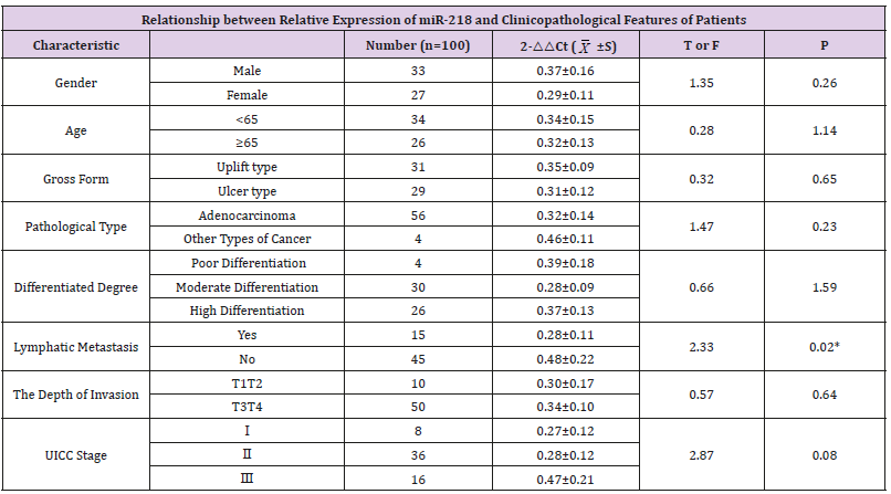
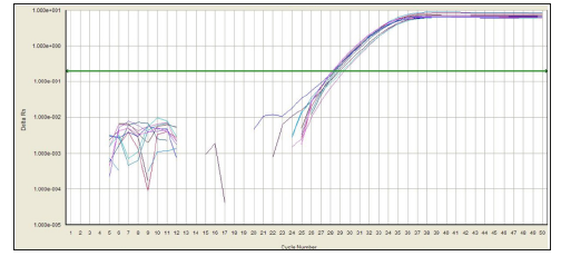
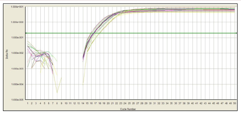
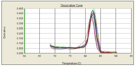
In this experiment, the amplification curve is concentrated, smooth, and has a stable plateau. The peak position is between 20 and 30 ct values (Figures 1 & 2), indicating the amplification efficiency, specificity and template of the primers in this experiment. The amount is more appropriate. The dissolution curve is concentrated in a single peak (Figures 3 & 4), indicating that the specificity of the test is good, and the product is single. In 60 cancer patients, the relative expression of miR-218 in 2 cases was >1, <1 in 58 cases and <0.5 in 4 cases. Relative expression was 0.3297 + 0.2998. Cancer and normal tissue of miRNAs - 218 express multiple 2- Δ Δ Ct difference (t = 17.320, P = 0.000). The relative expression level of miR-218 was not significantly different among all factors except lymph node metastasis (P>0.05), as shown in Table 1.
Table 1: Relationship between relative expression of miR-218 and clinicopathological features of patients.
Figure 1: Amplification curve of miR-218.
Figure 2: Amplification curve of U6.
Figure 3: Dissolution curve of miR-218.
Figure 4: Dissolution curve of U6.
By analysing more patients with colorectal cancer, miR- 218 expression was significantly decreased in colorectal cancer compared to non-tumor tissues. Low expression of miR-218 was also demonstrated in comparison of colon cancer cell lines with normal colon tissue [2]. The down-regulation of miR-218 also appeared in other cancers, including gastric cancer [3-4], glioma [5], nasopharyngeal cancer [6], lung cancer [7] and bladder cancer [8]. Human miR-218 contains two different genes (miR-218-1 and miR-218-2), which are processed to encode an identical mature sequence. MiR-218-1 and miR-218-2 were embedded in the introns of SLIT2 gene on chromosome 4p15.2 and SLIT3 gene on chromosome 5q35.1, respectively. 4p15.1 ~ 15.3 frequent absences can lead to colorectal cancer [9]. Therefore, heterozygous deletions in this region may be associated with down regulation of miR- 218 in colorectal cancer patients. On the other hand, SLIT family host genes are often inactivated in colorectal cancer through their promoter methylation [10]. Studies have found that the expression of miR-218 restores when the DNA methylation inhibitor 5-aza- 2 ‘-deoxycytidine is used in colorectal cancer cell lines. In nasopharyngeal carcinoma and oral squamous cell carcinoma, miR- 218 is down regulated by promoter methylation [11-12].
Lower Extremity Position Test: A Simple Quantitative Proprioception Assessment-https://biomedres01.blogspot.com/2020/11/lower-extremity-position-test-simple.html
More BJSTR Articles : https://biomedres01.blogspot.com


No comments:
Post a Comment
Note: Only a member of this blog may post a comment.