Anti-Radical Activity of Low Frequency and Low Amplitude PEMF
Introduction
If we expose biological tissue to Pulsed Electromagnetic Fields (PEMF) we can induce a measurable biological response and modulate inflammatory processes, promote bone repair, repair of skin wounds, even chronic ones, improve metabolic processes. Electromagnetic fields can interact with biological tissue inducing responses depending on their frequency and intensity. In the therapeutic field, the most promising results are obtained with the use of low intensity and very low frequency pulsed electromagnetic fields, or Extremely Low Frequency, indicated by the acronym PEMF LFLA. In fact, exposing cultures of fibroblasts and endothelial cells to PEMF LFLA, which vary during the stimulation process, we can quantitatively measure a reduction in the free radicals produced. Recent evidence has demonstrated that LPEMFs can prevent inflammation and oxidative stress as a non-invasive therapeutic method for various diseases. As an example, in 2019 a study [1] presented how, in an in vivo test, PEMF LFLA promote functional recovery following spinal cord injury, potentially by modulating inflammation, oxidative stress and HSP70. Furthermore, studies confirmed that PEMF LFLA can reduce ROS levels and enhance antioxidative stress responses in osteoblasts [2]. A polish clinical research [3] in 2018, attested that variable low frequency PEMF therapy meaningfully improved the overall condition of 57 patients through a decrease of oxidative stress markers while significantly affected positively their psychophysical abilities after stroke. In the present study it is presented a biophysical hypothesis of interaction between PEMF and biological working principle regarding the oxidative stress modulation PEMF derived, particularly in sport activities. Finally, we evidenced some results in vitro in fibroblast and endothelial cells and in vivo, in a case report on professional running athletes.
Biophysical Hypotheses Of Interaction between PEMF and Biological Tissues
There are several hypotheses of interaction between
electromagnetic fields and cellular structures. Among the various
hypotheses proposed in the scientific literature, the use of nonlinear
physical descriptive models, seem to be the best tools for
describing the induced biological phenomena. In particular, the
use of solutions of Schrödinger equation, relating to waves with
constant amplitude, called “solitons”, which will be better described
below, allows to explain, as a theoretical model, the effects of PEMFs
on the modulation of oxidative stress. The magnetic field can, in
fact, induce variations in electrical charges through the insulating
effect of the plasma membrane, acting on electrons, ions, radicals,
molecules and macromolecules.
The hypothesis of interaction of PEMF LFLA with cellular
structures includes (1):
1. Action on RNA polymerase and stimulation of cell replication
2. Action on the synthesis and hydrolysis of ATP
3. Effectiveness of oxidation-reduction reactions
Electro-magnetic fields can influence biological redox
processes. The electron transport chain, involved in the synthesis of
ATP and in respiration, is a process of oxygen reduction (by NADH
and FADH2) through electron transfer in the mitochondria (it is
the initial part of oxidative phosphorylation). The relatively small
mass molecules involved in the chain, such as cytochrome-c, can
move quite “easily” out of the mitochondrial membrane, carrying
electrons from donor to acceptor [4]. This electron transport can
be described using very complex physical theoretical modelling.
Trying to simplify, the various models consider electron donor
molecules loosely bound to “bridging” molecules, in turn loosely
bound to acceptor molecules. But if we consider high molecular
weight molecules such as the NADH-ubiquinone oxidoreductase
proteins and cytochrome c-oxidase, which are also involved in
the electron transport chain, these result to be practically fixed in
the inner mitochondrial membrane. Most of these proteins have
an alpha-helix conformation. When the electrons are released
from the donor molecule, they are transferred into the protein
and accompanied by a deformation of the endoplasmic reticulum
which forms a potential barrier that attracts the electron itself. A
relationship between protein conformation and formation of the
electron-lattice deformation bond is hypothesized.
The charges then propagate, accompanied by a deformation
of the lattice. Mathematically, through the solutions of non-linear
equations, it is possible to describe this propagation which assumes
a waveform with particular characteristics, such as the amplitude
that remains constant. We will see that this wave has been called
“soliton”. To demonstrate this result, the ion-resonance frequency
relationship can be inserted into the solution of the non-linear
Schrödinger equation, defined in quantum mechanics [5]. If we
consider the presence of potential U(x,t) the formula of the equation
can be written as follows:

Cyclotron ion resonance is a phenomenon related to moving ions immersed in a magnetic field and is based on the Lorentz force. The magnetic field acts on electronically charged elements such as ions (eg Na +, K +, Ca ++) and electrons, certainly available in biological systems. The magnetic field can act on the movement of these electric charges through a force called Lorentz and defined by

where the contribution of the electric field E which acts on the charge q, and the contribution of the magnetic field of intensity B, which acts on the same charge as a function of its speed v, determines the intensity of the force F. Thanks to the Lorentz force, a free ion immersed in a magnetic field of intensity B, moves on a circular trajectory with an angular velocity ω, dependent not only on the intensity of the magnetic field but also on its own charge and mass. Using the information of the Lorentz force and the circular trajectory, the following formula can be obtained:

If we insert this frequency value into Schrödinger’s equation
and solve it, the solution obtained is the wave function of the
electron ψ (x,t ) .
More correctly, its square provides information on the
probability of finding the electron when trying to measure its
position. Applied to the electron transport chain, the wave function
provides information on the position of the particle along the protein
lattice. Considering an alpha polypeptide chain, the solution of
this equation is a particular wave, which indefinitely maintains its
amplitude. In the literature this wave has been descripted through
the shape of a “soliton”.

The soliton is a solitary wave that keeps its amplitude
constant and represents the “module” for transporting energy
and information at the cellular level. With the same solution we
can calculate the deformation of the lattice (chain) in the “soliton”
state, which is proportional to the probability of the presence of the
electron (x). From the mathematical demonstration we obtain that
the electron and the deformation of the lattice together, propagate
along the polypeptide chain, with constant speed, from a donor
molecule to an acceptor molecule, as a localized wave. This wave is
exceptionally stable, able to propagate along macroscopic distances,
thanks to the combination of low energy dispersion and non-linear
phenomena (non-linear nature of its formation). The oscillatory
character of soliton propagation with frequency given by Equation
1 is accompanied by the propagation of the local deformation of the
polypeptide chain.
At a “macroscopic” level, solitons are well known and used in
some fields of application. For example, in optical fibre transport
networks, the non-monochromatic light, due to the Kerr effect,
can propagate without changing over time. Tsunamis are common
in seismology, generated by submarine earthquakes, capable
of spreading for kilometres without dispersing energy, before
impacting the coasts, with all their destructive force. They are
therefore waves that do not lose their energy and their intrinsic
characteristics by propagating and this is due to the combination of
dispersion and non-linear effects, which interact by cancelling each
other. The name “soliton” is due to their characteristic of remaining
unchanged in the event of reciprocal interaction: two soliton
waves that collide do not add up their effects. The hypothesis
that some biological phenomena are based on the presence of
solitons is due to Alexander Davydov who in 1979 and then in
1985 hypothesized their existence in alpha polypeptide chains [4].
Davidov demonstrated, mathematically, that the energy from ATP
hydrolysis could be stored as a vibration of the amide group of the
alpha helix protein, through a non-linear interaction that traps, the
energy into a soliton [6,7]. The energy comes from the hydrolysis
of ATP, as widely known in biology, but this is stored in the form
of mechanical vibration of the amide, with constant amplitude.
The subsequent propagation of this energy can explain the muscle
contraction [8] (Figure 1). Over the years, scientific literature has
shown, mathematically, how the solitons mechanism was a good
solution to explain biological phenomena. It therefore becomes
clear how PEMF LFLA can interact with these mechanisms,
promoting biological phenomena.
Figure 1: Alexander Davydov was the first to hypothesize the action of solitons in biological systems.
Biological Working Principle of ROS Regulation with PEMF Exposure
Both in the therapeutic and sports fields, an important effect
of PEMF LFLA is the reduction or, more correctly, the regulation
of oxidative stress. This plays an important role in numerous
pathological conditions on an inflammatory basis, involving
the cardiovascular, neurological, metabolic and respiratory
compartments. In the respiratory tree, free radicals cause epithelial
and vascular necrosis, connective tissue degradation and activation
of pro-inflammatory factors [9]. Sports-related oxidative stress
slows post-workout recovery, increases fatigue and the risk of
myocyte injury. ROS free radicals are produced during physiological
chemical reactions that use oxygen. These molecules are particularly
reactive and unstable due to at least one unpaired electron in their
outer orbital. To reach an electromagnetic equilibrium state, they
try to “recover” that electron binding other atoms and leading to
new and further unstable molecules. Modulated PEMF LF LA can
reduce the average “duration” of the oxidative free radical, limiting
its damage action, in particular during inflammatory processes,
both acute and chronic, which limit the physiological healing
processes or sports recovery. The “spin chemistry”, through the
“radical pair mechanism”, can describe the mode of action of the
low intensity magnetic field with chemical reactions [10].
During an oxidative reaction, radical pairs are formed, therefore,
as known, with “unpaired” electrons. These electrons can be
“parallel” or “antiparallel”. In the first case the radical couple will be
in the electronic “triplet” state, while in the second case the couple
will be in the “singlet” state [11]. During the chemical reaction,
with the formation of the radical pair, there is an interconversion
between singlet and triplet states. This reaction influences the
speed and the course of the chemical process and can be influenced
by applied magnetic fields, in particular if they have low energy and
variable frequency. The PEMF LFLA fields therefore promote the
transition to a more stable state, that is the “triplet” state, reducing
the “average life”, that is the duration, of the free radical [12].
In vitro tests
In order to demonstrate the ROS-reducing action, PEMF LF LA was applied to in vitro cultures of fibroblasts and endothelial cells [5]. By means of a solenoid with 54 coils and a generator of sinusoidal electrical signals that allowed the combination through DDS of several sinusoids at different frequencies and above all, to make the frequencies vary during the treatment, impulsive e.m. fields have been generated. at frequencies that can be varied in the range 0-300Hz and with average intensities of 100 μT. In conditions of oxidative stress, free radicals accumulate, but by exposing cell cultures to modulated pulsed electromagnetic fields, it is possible to limit their production, or, more correctly, reduce the duration of activity of ROS by reducing their quantity overall. The following figure (Figure 2) shows the comparison of the quantities of ROS, measured by the Oxyselect kit in cultures of fibroblasts and endothelial cells [5], exposed to the described LF LAPEMF 20 minutes/day for 21 days [13]. The curves show the progressive variations of ROS species on a daily basis: in fibroblasts a reduction is observed, compared to the control, starting from the 8th day, while in endothelial cells the reduction of ROS is evident from the first exposure to PEMF LFLA 0-300Hz, 100uT.
Figure 2: Quantity of ROS species in fibroblast (left) and endothelial (right) cells vs treatment days (20min / d): green line with squares represents the treated cells, while the red line with rhombuses the control cultures.
Clinical Test Report on Athletes
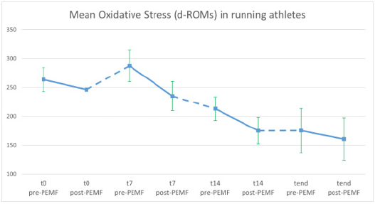
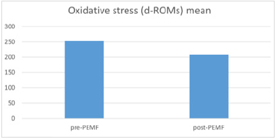
An explorative clinical test report was performed in order to evaluate the ROS influence as a systemic effect after low intensity and low frequency PEMF exposure. The PEMF emitting systems was composed by a mattress with three pairs of solenoids at increasing numbers of coils (18, 36, 50). The coils are connected to a generator which supplies the therapy as per in vitro tests (variable frequencies 0-300 Hz and variable intensity 100 μT mean). We measured systemic ROS levels of n.5 professional athletes with the use of a portable system (FRAS 5, H&D S.r.l. – Italy) before and after exposure with PEMF at different timepoints during their training session. A small amount of capillary blood was taken from the fingertip and analysed through photometer which determines the blood concentration of ROM (Reactive Oxygen Metabolites = Free radicals) in U CARR (10). Patients undergo a 30-minute PEMF exposure lying on the mattress with solenoids once a day. At time 0, 7, 14 and end time (21 days) the analysis of the values of free radicals (d-ROMs) were carried out, before and after a treatment session with PEMF, and then athletes continued the treatment at home. The points in the above figure (Figure 3) showed a decrease of the measurement of systemic ROS after 30 minutes of exposure with PEMF in every session resulting in an overall decrease at the end of the study. This is confirmed by the mean between the measurement before and after PEMF treatment throughout the study in five professional running athletes (Figure 4).
Figure 3: Mean of d-ROMs in U CARR in capillary blood of 5 athletes before (pre-PEMF) and after (post-PEMF) exposure with PEMF treatment at start of treatment (t0) and after 7, 14 and 21 days (tend). In green the standard deviation.
Figure 4: Overall Mean of d-ROMs in U CARR in 5 athletes before (pre-PEMF) and after (post-PEMF) exposure with PEMF treatment of the 4 sessions.
Discussion
LFLA PEMFs are able to interact with biological tissue and modify its responses, for example by reducing free radicals and consequently their action in inflammatory processes. The theoretical hypothesis is based on the modulation of soliton waves capable of acting on cellular elements with an electric charge, in the processes of cellular respiration, realizing the transport of electrons, and promoting the formation of stable states and reducing the duration of action of the free radical. Initial evidence has been found in vitro in fibroblast and endothelial cells, where the exposure to PEMF resulted in a decrease of ROS levels. Clinical exploratory test report on professional athletes showed a decrease of systemic ROS levels after PEMF exposure. LFLA PEMF showed capability to reduce ROS level in vitro and in vivo, but further studies need to be implemented in a wide population in order to confirm this first experimental results.
For more
Articles on : https://biomedres01.blogspot.com/


No comments:
Post a Comment
Note: Only a member of this blog may post a comment.