The Effects of Concurrent Training during Cardiac Rehabilitation on Plasma MMP-9 and TIMP-1 Levels in Myocardial Ischemic Patients
Introduction
Cardiac ischemia is considered as one of the deadliest heart
diseases. According to the reports from American Heart Association,
one third of cardiac patients suffer from ischemic heart ailment
[1]. Several genomic and environmental factors including but
not limited to family history, gender, age, smoking, hypertension,
diabetes, hyperlipidemia, and lack of physical activity can potentially
contribute to atherosclerosis and subsequently ischemic heart
disease [2,3]. Atherosclerosis, a chronic inflammatory disease, is
characterized as the accumulation of fat and other substances on
the inner surface of artery walls [4]. In the atherosclerotic situation,
due to the deficiency of blood flow in the coronary arteries, an
acute blockage occurs which is defined as a heart attack [5]. With
the progression of atherosclerosis, the smooth muscle cells move
toward the intima in response to growth factors released by active
macrophages and endothelial cells. Active macrophages weaken the
extracellular matrix and support the platelet fibrosis by producing
matrix metalloproteinases (MMPs) suggesting a prominent role
in the process of atherosclerosis and rupturing the artery walls
[6]. Matrix metalloproteinases (MMPs), which are a large group
of protease enzymes, are responsible for the breaking down of
extracellular matrix.
To date, 26 members of MMP family have been identified, and
among them, gelatinase-B (or MMP-9) has a higher activity than
other members [7] with a well-documented potential in developing
cardiovascular disease in human populations [8]. Based on the
evidence, myocardium expression of MMP-9 is increased in the
patients with coronary artery disease [9,10]. However, due to the
damaging role of MMPs, their activity is strongly regulating by four
tissue inhibitors (TIMPs) namely TIMP-1, TIMP-2, TIMP-3, and
TIMP-4 which can control the detrimental activities of MPPs in
vertebrates [11]. Among the TIMP family, it has been reported that
TIMP-1 is a potent inhibitor of many MMPs including MMP-9 [12]
and play a key role in the structure and function of myocardium
ae well as the control of extracellular matrix proteolysis in
cardiovascular system [12]. Recurrence of cardiac ischemia
and its unfavorable consequences are a major concern among
cardiac ischemic patients as well as cardiovascular specialists [8].
Nowadays, with developing clinical centers, medical strategies,
and patients’ knowledge in diagnosing early signs of heart disease
the rate of mortality has been clearly decreased; however, some of
these patients with the acute symptoms have been hospitalized for
the second and third time and some of them still may have died
[13].
In addition to pharmacological treatments and implementing
medical interventions on coronary arteries, cardiac rehabilitation
program has been well-documented in controlling the risk factors,
improving life quality, and reducing the mortality rate [14]. The
cardiac rehabilitation program is designed to limit the physiological
and psychological consequences of cardiovascular disease, reduce
the risk of sudden death or stroke, control cardiac symptoms, and
decline the atherosclerosis [15]. Physical exercise is frequently
used as a non-pharmacological and supplemental method in the
process of medical treatment for a variety of reasons [16]. As an
incentive method, physical exercise can be used during cardiac
rehabilitation programs due to its low cost and attractiveness
[17]. The type of physical exercise used in cardiac rehabilitation
programs is generally aerobic in forms of walking, running, or
cycling [18]. However, it has been recently reported that a welldesigned
resistance training program accompanied by aerobic
exercise can be considered as a helpful strategy in rehabilitation
programs. In this regard, the American Heart Association has also
recommended that resistance training be performed twice a week
in cardiac rehabilitation programs [18].
According to the recent scientific reports, the implementation
of physical exercise in rehabilitation programs can lead to effective
outcomes on some clinical indicators involved in the occurrence
of atherosclerosis [19]. In a study examining the effect of cardiac
rehabilitation on the atherosclerosis biomarkers in patients with
cardiac ischemia, the authors reported that performing physical
exercise during cardiac rehabilitation programs will prevent reoccurrence
of cardiac ischemia [20]. However, due to the increasing
prevalence of ischemic heart disease [1] and considering the
importance role of MMPs particularly MMP-9 in the formation
and stabilization of atherosclerotic plaque, few studies have yet
examined the effects of a well-planed physical exercise during
cardiac rehabilitation on the levels of metalloproteinases and
their tissue inhibitors in patients with cardiac ischemia [21,22].
Therefore, the need for a comprehensive investigation in this
promising area is clearly evident and the present study is designed
to examine this imperative issue.
Materials and Methods
The present study is an experimental controlled clinical trial.
Sixteen cardiovascular patients with atherosclerotic symptoms who
were referred to a cardiac hospital in Mashhad, Iran were selected
and randomly assigned to an experimental (n = 8) or control (n = 8)
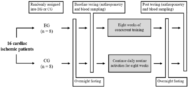
group. Scheme of the study design is shown in Figure 1. Before the
commencement of the study, patients in both groups were monitored
by a cardiologist and were reported to have symptoms of coronary
heart disease. For each patient, a file including demographic
information, history, clinical cardiac reports, and anthropometric
data was provided. At the beginning of the rehabilitation program,
an initial assessment of cardiac function was conducted using
echocardiographic devices. During the study, participants in both
groups followed the prescribed medications and diets (with a
particular amount of calorie and macronutrients per kilogram of
body weight for all participants) which were provided by a dietitian and
a cardiologist. The patients in experimental group completed
an eight-week moderate concurrent exercise program with 40-60%
of their one maximum repetition.
According to the American Heart Association guidelines, the
patient’s exercise intensity was monitored to be between 60 and
80% of their maximum heart rate [23] using the individualized
electrocardiograms connecting to each participant during exercise
sessions. Moreover, Borg Rating of Perceived Exertion (RPE) scale
was also used to a further control of the exercise intensity, and the
patients in experimental group were asked to keep their activity
intensity between level 11 (relatively light) and level 13 (somewhat
difficult) [24]. The concurrent training protocol is shown in Table
1. Before the commencement of training sessions, participants in
experimental group were informed and introduced to devices and
exercises in one session and then practiced three sessions per
week for eight weeks. Each training session lasted about one hour
performing mixed aerobic and anaerobic exercises using treadmills,
bikes, ergometers, physio balls, and light weights. At the starting of
each raining sessions 5 minutes of warm-up, 5 minutes of walking
on treadmill, 5 minutes of bike pedaling, and 5 minutes of upperbody
ergometer pedaling were done with three minutes of inactive
rest between each aerobic exercise.
After 5 minutes of rest, the individuals gradually performed
the following workouts with 10 repetitions in three sets in the
initial training sessions and to 15 repetitions in the advanced
sessions. The workouts consisted of squat with a physio ball,
shoulder flexion, shoulder abduction, elbow flexion, hip flexion,
hip abduction, ankle plantar flexion, and ankle dorsi flexion. The
workouts were initially performed using the patient’s own body
weight or limb weight, and gradually improved using therabands
and light weights [25]. A careful supervision was applied during
the training sessions, and the patients were constantly questioned
about the amount of pressure based on the Borg scale. If the value
of Borg scale was reported below 11 or above 13, the participant
was asked to increase or decrease the effort, respectively.
Laboratory Methods: Fasting 10cc blood samples were
obtained 48 hours before the first training session and 48 hours
after the last training session from the participants’ antecubital
vein in both groups. Blood samples were collected in lavendertop
tubes containing anti-coagulant EDTA, and then were carried
to laboratory for plasma separation by 3000rpm centrifuging
for 10 minutes. Subsequently, plasma samples were frozen and
stored at -80°C for further analysis. Plasma MMP-9 and TIMP-1
concentrations were assessed by Enzyme-Linked Immunosorbent
Assay (ELISA) method using Awareness Stat Fax 2100 device
according to the instructions of the ELISA kits (R&D Systems,
Minneapolis, MN, USA).
Results
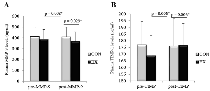
Data distribution was normal according to the results from Shapiro Wilk test. The baseline characteristics of the participants are shown in Table 2. The independent T-test showed a significant decrease in plasma MMP-9 concentration (t = 2.431; p = 0.029) and a significant increase in TIMP-1 plasma levels (t = 3.202; p = 0.006) following the exercise intervention in the exercise group compared to the control group. According to the correlated T-test, a significant within-group decrease in MMP-9 levels (t = 0.695; p = 0.008) and a significant increase in plasma TIMP-1 concentrations (t = 3.964; p = 0.005) were observed in the experimental group, while within-group changes in MMP-9 and TIMP-1 levels were not significant in the control group. (t = 0.21; p = 0.838 and t = 0.46; p = 0.66, respectively) (Figure 2).
Table 2: Baseline characteristics of the participants in experimental (n = 8) and control (n = 8) groups (mean ± SD).
Figure 2: The average changes in plasma levels of MMP-9 (part A) and TIMP-1 (part B) between per- and post-test values in experimental (EX) and control (CON) groups.
Discussion
Present study showed that after eight weeks of moderate
physical exercise during the cardiac rehabilitation period, MMP-
9 levels decreased and TIMP-1 levels increased, and significant
within-group changes in MMP-9 and TIMP-1 levels were observed
in the experimental group. To date, the investigations assessing
the effects of exercise on MMPs and TIMPs levels have often
been conducted in healthy, obese, or diabetic populations as well
as animal models. Seydanlou and Farzanegi in 2014 reported a
significant 20% decrease in MMP-2 levels and a significant 26% increase
in TIMP-1 levels of overweight individuals following eight
weeks of Pilates training [26]. Consistent with our results, Kadoglou,
et al. in 2013 found a significant increase in TIMP-1 concentrations
following six weeks of treadmill running in animal models [27],
similar to the results of Koskkinen’s study but in humans [28].
Although the effectiveness of physical exercise on MMP-9 and
TIMP-1 levels are well documented in the literature, limited studies
have reported no change in the aforementioned variables following
different exercise interventions. Previously, Mackey, et al. reported
that a 10-kilometer road and water running has no effect on MMP-2
and MMP-9 levels in young men [29].
Moreover, according to the results of a study by Hoier, et al.,
eight weeks of cycling training with the intensity of 60% maximum
oxygen consumption did not affect the TIMP-1 levels in healthy
men [30]. However, these inconsistent findings might be justified
by different training methods, varied participants’ characteristics,
and different sampling methods (muscle biopsy vs blood sample)
used in the aforesaid investigations. Metalloproteinase matrix
enzymes are believed to alter the formation of the cardiovascular
matrix during natural biological processes [31]. However, in
pathophysiological processes of various diseases, the expression
and activity of these type of proteolytic enzymes are increased
due to the increased secretion of proinflammatory cytokines
leading to breakdown of several collagens and gelatinases, as well
as detrimental effects on microanatomical tissue structures. As a
result, exacerbation of inflammatory state and emerging of various
heart and vascular diseases might occur with advancing time
[32,33]. In a study examining the role of matrix metalloproteinase
1, 2, 3, and 9 in acute myocardial infarction, the authors reported
that the plasma concentrations of various MMPs in patients with
myocardial infarction were significantly increased [8].
They concluded MMP-9 may play a prominent physiological
and pathological role during the stage of myocardial infarction to
heart failure, more especially considering the fact that improper
vascular redisposition of MMP-9 may promote atherosclerosis via
weakening of atherosclerotic plaques [34]. It seems the potential
mechanisms for reducing exercise-induced MMP-9 levels during
cardiac rehabilitation is the changes in the levels of monocyte
chemoattractant protein-1 (MCP-1) [35]. MCP-1 attracts monocytes
to inflammatory sites located in the vascular subendothelial space.
These monocytes are able to differentiate into macrophages and
be converted to foam cells via absorbing oxidized low-density
lipoproteins (Ox-LDL), suggesting an important role for MCP-1 in
the pathogenesis of atherosclerosis [36]. The inhibition of MCP-
1 by relevant inhibitors prevents plaque inflammation and stops
the rupture of disposed plaques [37]. On a better note, MCP-1 in
myocytes and smooth vascular cells stimulates the expression of
MMP enzymes and consequently induces inflammatory cytokines
and MMP enzymes in cardiac myocytes [38].
Although MCP-1 levels were not measured in the present study,
several studies have reported a reduction in MCP-1 following
exercise suggesting a beneficial effect of physical exercise on heart
patients. Thus, MCP-1 reduction is likely links to decreased plasma
MMP-9 levels following regular exercise [39]. The activity of MMP
inhibitors and the ratio of MMPs to TIMPs are as important as the
secretion of tissue MMPs. Under normal conditions, a physiological
balance between MMPs and TIMPs is stablished, and the
extracellular matrix breakdown and synthesis is well-adjusted. Any
disease or mechanical stress that results in decreased immune cells,
increased inflammation, secretion of proinflammatory cytokines,
and the activity of MMPs, consequently triggers the immediate
inhibitory response particularly activation of TIMPs [40]. Hence,
the inhibition of MMP proteolytic activity has been suggested as a
therapeutic approach in various heart diseases [41-43].
Conclusion
In the present study, an inverse relationship between the bioactivity of MMP-9 and TIMP-1 was observed following eight weeks of exercise intervention in ischemic cardiac patients. Considering the favorable effects of physical exercise on cardiac rehabilitation, it can be suggested that patients with cardiac ischemia can accelerate their recovery process and reduce the risk of stroke reoccurrence by doing moderate intensity of physical exercise at least three time per week.
For more
Articles on : https://biomedres01.blogspot.com/


No comments:
Post a Comment
Note: Only a member of this blog may post a comment.