Virtual Rapid Sensing of Covid-19 Virus Antibodies in Patients Blood by Using Platinized Antigen Linking Graphene Probe
Introduction
In December 2019, a cluster of atypical pneumonia cases was detected in Wuhan, China, which soon spread throughout China before Chinese Lunar New Year in January 25, 2020. Outdoor movement restrictions were also adopted in a number of wide cities. However it was far too late. Here the novel coronavirus was named COVID-19 by the World Health Organization (WHO) on February 11, 2020. Later the COVID-19 epidemic was declared a public health emergency of international concern by WHO on January 30, 2020. Also later, on March 11, 2020 it was declared a pandemic by the WHO. On March 29, 2021, more than 126.89 million people have been confirmed to be infected (across over 223 countries, areas or territories), of which more than 2.78 million people died (WHO, 2020b) [1]. This is now pushing the whole of humanity into a biological warfare against viruses. Recent virus detection methods depend on the titrimetric PCR amplifications such as hemagglutination inhibition assay of the microneutralization [2], fluorescence spectrometric absorption method [3], photometric UV-Vis spectroscopy and electron microscopy [4] and others. Here conventional testing methods require approximately 1~8 days by virus culture, DNA amplification, electrophoresis separation and neutralization titration spectroscopic analysis. However, electrochemical in vivo or in vitro trace ion [5] and [6] cell [7,8] detection is fast, and only a few minutes are required. Diagnostic circuit fabrications are simple, so for this reason, we have developed modified probe sensing skills such as micro implementation [9] Diagnostic carcinoembryonic antigen tumor marker [10] detection, Pico molar ranges [11] working probe infrared photo diode electrodes [12,13] for in vivo cell [14] or human fluid [15] non treated tissue direct assay [16] and macro [17] synthesis [18] Human Blood Plasma [19] sensing ect, also which sensing circuit can be interfaced to telemetric WiFi wearable diagnostics for covid virus detection. Here developed results can replace the existing complex WHO method, such as PCR DNA amplification, Fluorescence Absorption Analysis, or chromatographic separation.
WHO Standard Diagnostic Protocol Comparison
Conventional WHO diagnostic protocols of PCR DNA amplication titrimetric methods is time consuming and replicated sophisticated skills are required. Also highly trained master’s and doctoral technicians are required: for example, Real-time RT-PCR assays for Eurasian H7HA standard protocols shown as follows
WHO Protocol 2
Real-time RT-PCR assays for Eurasian H7HA shown here.
Materials Required
1. QIAamp Viral RNA Mini Kit (QIAGEN®, Cat. No.52906) or equivalent extraction kit
2. ThermoFisher Fast Virus 1-step Master Mix (Applied Biosystems®, Cat. No. 44-2)
3. Ethanol (96–100%)
4. Microcentrifuge (adjustable, up to 13 000rpm)
5. Benchtop centrifuge with 96-well plate adaptor
6. Adjustable pipettes (10, 20, 100, 200μl).
7. Sterile, RNase-free pipette tips with aerosol barrier.
8. Vortexer
9. MicroAmp Fast Optical96-well reaction plate(Life Technologies, Part No.4346906)
10. MicroAmp optical adhesive film (Life Technologies, Part No. 4311971)
11. Real-time PCR system (ViiA™ 7 Real-Time PCR System)
12. Positive control (Available from HKU, e-mail: llmpoon@hkucc. hku.hk)
13. Primer and probe set.
Type/subtype
Influenza A (H7Nx). Gene:HA.
Name and Sequence
HA H7-1603F: TTTAGCTTCGGGGCATCATGTTT
H7-1674R: CAAATAGTGCACCGCATGTTTCCA
H7-1646P: FAM-TGGGCCTTGTCTTCATATGTGTAAA-MGB
Procedure from reference [20]
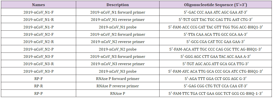
For more detailed examples of Covid 19 primers and probes sequences shown here. also which sequence was used to antigen N1~N3 mixing immobilized working probe fabrication. At this work. Here Table 1 shows the coronavirus disease 2019.molecular DNA sequence [21]. This DNA was used for the working probe mixed antigen immobiliz. The following images showing synthetic polyma in vitro film and sensing probe schimetic fabrication.
Materials and Methods Working Probe
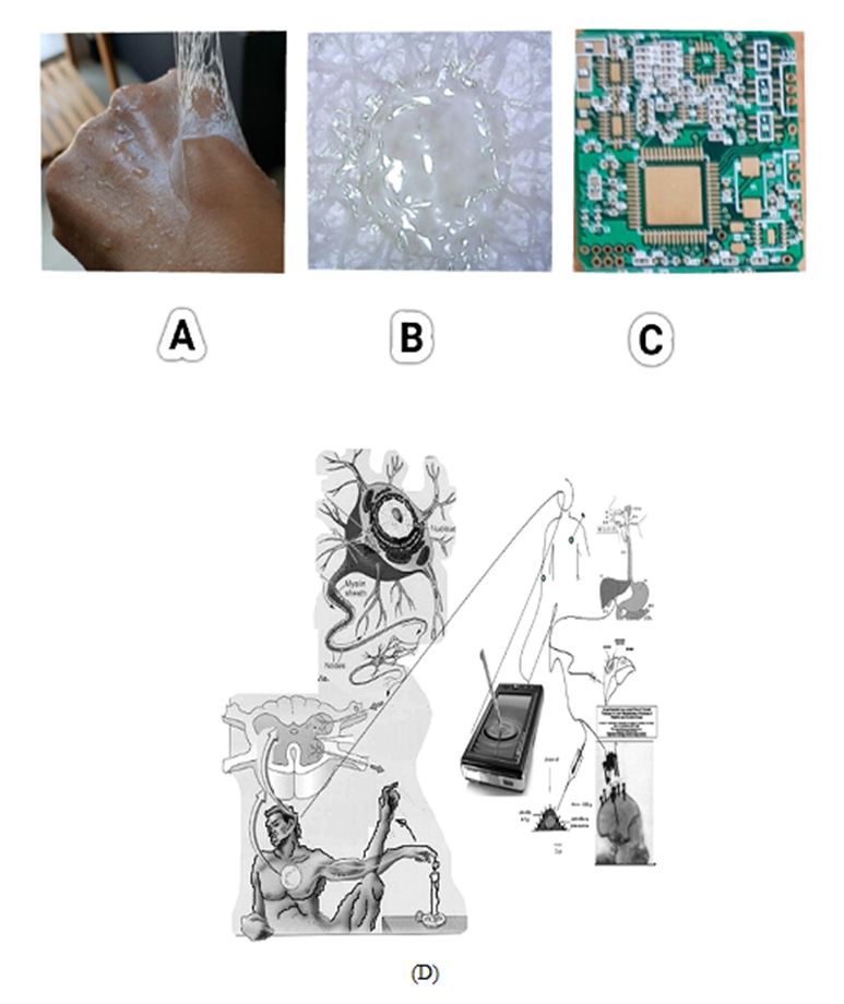
(Figure 1A) A image is shown for skin tattoo coated conduction film made from synthesizing water-dispersion resin with the following characteristics: Softness, Wear-resistance, life extension, Skin protection, Conductivity and Transparency. Image B is a tattoo working electrode at the in vitro skin muscle electrode. Image C is voltammetric bio-electroanalyzer workstation with Wi-Fi telecommunications. It can be used to wearable diagnostic circuit for virus detection. Here in three electrode systems of counter and reference electrode was made like image B. However, working electrode fabrication is shown here, and the probe binder was used by synthesizing an aqueous dispersion resin. The resin paste was made with graphene nanopowder, and covid antigen, and platinum catalys were mixed with conductivity resin. Here the antibody antigen redox titration potential was obtained to reductions of -0.14V, 0.26V anodic scanning and - 0.9V oxidation peak currents was obtained during cathodic scanning. Analytical in vitro WiFi application is shown at (D).
Figure 1: (A) Skin tattoo micro rasin coating film.
(B) In vitro muscle skin tattoo working probe.
(C) Voltammetric Wi-Fi wearable coin size workstation.
(D) Virtual diagnostic in vitro schematic diagram of the working probe redox voltammetric ionic reaction for antigen immobilized on a graphene nanotube synthesize antibody antigen titration image.
Electrochemical Analyzer
Diagnostic circuit was used for the bioelectrochemical analyzer-2. voltammetric and our system, which can be designed to 5Cm×3Cm×60mm like physical coin size with telemetric WiFi control wearable workstation, potential windows of 2.0V~-2.0V redox potential windows, scanning. The current amplification of 1.0×10⁻³A~1.0×10⁻⁹A pico range and cyclic voltammetry redox, stripping voltammetry and chronoamperometry 2D visual control programs were used, power of USB interfaced ±2.5V strength was used.
Working Electrode Preparation
The diagnostic working probe was made by maxing a ratio of the graphene nano paste 2.0 g, mineral oil 0.5 g, Water-dispersible synthetic resin 0.5 g, platinum standard solution 1000 ppm 0.5 g. Paste was mixed completely during 20 minutes. The paste was coated on the graphite crystal load of 0.5 mm diameter 30 mm length, then connected to voltammetric working electrode terminal by using 0.35 mm pure copper enamel insulation coating wire, counter and reference electrode was made with the same method using graphite crystal load. The circuits are usable for wearable tattoo assay such as In-vivo or in vitro and feeling sensing analysis.
Results
Results and Discussion
Voltammetric analytical optimum para conditions were searched in PBS 5 mL electrolyte microtube conical rack, with 0.6 mL covid virus antigen spiking, at room temperature. The cyclic voltammetric antigen and antibody redox ionic voltammograms were searched.
Cyclic Voltammetric Optimization
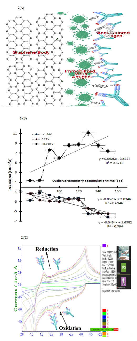
Cyclic potential scanning is related to covid 19 virus ionic redox activation. For this purpose, the cyclic scan rate was searched from 0.2mv/sec~0.9mv/sec eight points in 0.9 mL antibody contained PBS buffer, 30 sec stirring convection with magnetic bar, only changed for scan rate variations. The real voltammogrms shown at Figure 2(A) is antigen antibody titration working image. (B) is, 0.2mv/sec scan rate oxidation peak obtained two site at -0.14V peak high of (4.322×10⁻⁵A) and 0.14V peak high of (3.46×10⁻⁵A) anodic current obtained then reduction peak of -0.9V peak high of (10.4×10⁻⁵A) cathodic obtained, which anodic and cathodic linear statistic 3rd equations shown there slope gradient, intercept and relative standard deviations, here in -0.14V potentials current linear increased, dx/dy=-31.782, y intersect is 3.4125, R2=0.8351 obtained. Other statistic equations not shown here. Figure (C) is reduction potentials of two peaks and oxidation of one linear equation obtained, least square linear equation and relative statistic results not shown in this figure, and the accumulation time increase. The sensitivity increase obtain, here qualitative potentials windows was used to square wave stripping voltammetry.
Discussion
Stripping Voltammetric Optimization
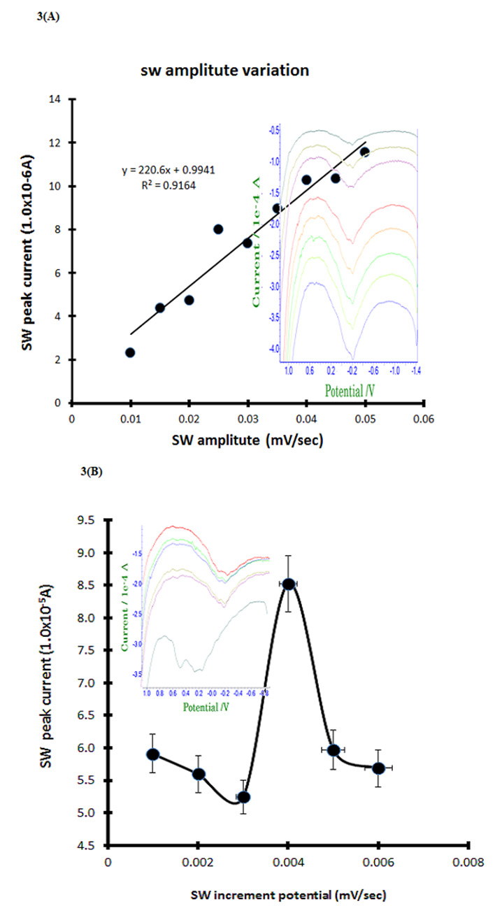
Cyclic voltammetric antigen and antibody anodic or cathodic ionization potentials were applied to sensitive stripping voltammetry. Here stripping accumulated amplifications are increasing to the detection limit, so anodic and cathodic scanning accumulation time variations were examined in 0.9 mL covid 19 antibody containing the PBS electrolyte. Stripping optimum amplified variations shown in Figure 3A, at the first point of 0.01 mV/Sec is 2.321×10⁻⁵A obtained in -0.2V potential windows anodic scanning, then continue variations performed during 10 steps by same methods, here final step is 0.05 mV/Sec 11.54×10⁻⁵A obtained on the linear curve. The least square linear equation is shown inserted in picture of Y=220.06x+0.9941, slop sensitivity is dx/ dy=220.06, intercept is 0.9941 and the relative standard deviation is R2=0.9164 obtained, at this para conditions square wave increments variation was examined from 0.001 mV/Sec to 0.006 mV/Sec with six steps, results shown in Figure 3B voltammograns and linear statistic equation, here in 0.004 mV/Sec is big current of 8.524×10⁻⁵A obtained, thus 0.05 V/Sec amplitude and 0.006 V/Sec increments are fixed. Under these conditions, diagnostic concentration effects were examined.
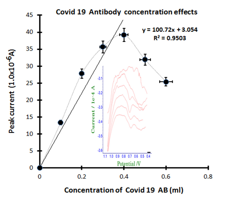
Figure 4 is analytical application effects for patients’ blood extracted antibody standard spike. The optimized final para conditions, diagnostic real antibody was examined in the PBS buffer electrolyte 8 mL volume. The first peak of electrolyte blank stripping is non signal appeared, then the 0.1 mL antibody spike was obtained to anodic of 0.7V oxidation current for 13.37×10⁻⁶A. In this figure, the inserted voltammograms shown slowly increased the sharp width, then continued spiking of 0.2, 0.3, 0.4, 0.5 and 0.6 mL the same conditions were added. Here the peak current 27.78×10⁻⁶A, 35.65×10⁻⁶A, 39.21×10⁻⁶A, linearly increased, then 31.92×10⁻⁶A, 25.37×10⁻⁶A decreased, which linear equations of dx/ dy=100.72, Y intercept is 3.054, the relative standard is R2=0.9504, which statistic results can be usable for real patients’ blood assay, anytime and anywhere immediately with self-detection. Thus, a more simplified tattoo wearable sensing circuit was fabricated, which can be used to telemetric remote control.
Figure 2: (A) Cyclic voltammetric scan rate variations for 0.2, 0.3, 0.4, 0.5, 0.6, 0.7, 0.8, 0.9 mV/Sec, insert voltammograms shown reductions of -0.14V, 0.26V and -0.9V oxidation peak currents with 0.9 mL antigen spike in 3mL PBS electrolyte.
(B) Cyclic voltammetric accumulation time variations for 70, 80, 90, 100, 110, 120, 130, 140, 150 sec at (A) condition.
(C) Virtual real time voltammograms ionic imager.
Figure 3: (A) Square wave stripping amplitude variations for 0.01, 0.015, 0.02, 0.025, 0.03, 0.935, 0.04, 0.045, 0.05 V/Sec in PBS electrolyte 3mL, with 0.9mL Covid 19 antigen spike.
(B) Square wave increments potential variations for 0.001, 0.092, 0.03, 0.094, 0.005, 0.06 V/Sec of (A) electrolyte conditions.
Figure 4: Concentration effect of Covid-19 antibody for 0.1mL, 0.2mL, 0.3mL, 0 4mL, 0.5mL, 0.6mL standard spike, during stripping optimum para conditions, accumulation time was 60 sec string anodic -2.0V~-2.0V in PBS 8 mL buffer electrolyte.
Conclusion
Diagnostic optimum results be attained in 60 seconds accumulation time only. During the anodic scanning, stripping peak potentials of 0.7V oxidation current was obtained of antibody antigen ionic transfer redox strength. Electrolyte buffer solution needed 5 mL PBS only and didn’t require any titrate or separation solutions. Detection systems used to Wi-Fi telemetric voltamnetric workstations of wearable patch type simple circuit of -2.0V ~ -2.0V potential scanning cyclic amplifier, working electrode is tattoo synthetic art painting film sensors of three electrode. The current range was 1.0×10⁻⁴A ~ 1.0×10⁻⁵A. The developed diagnostic results be used for virus detection and in any other field requiring human computer interfaced wearable tattoo bioassay and in vivo remote telemetric treatments.
For more Articles on: https://biomedres01.blogspot.com/


No comments:
Post a Comment
Note: Only a member of this blog may post a comment.